

130
7 Reactive μCP on Ultrathin Block Copolymer Films …
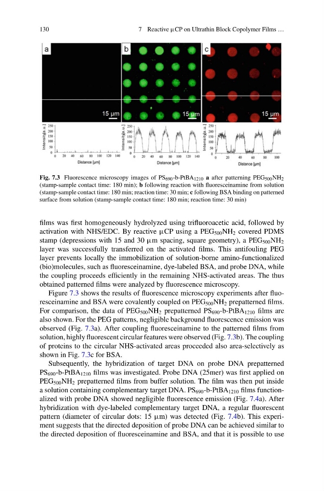
Fig. 7.3 Fluorescence microscopy images of PS 690 -b-PtBA 1210 a after patterning PEG 500 NH 2
(stamp-sample contact time: 180 min); b following reaction with fluoresceinamine from solution
(stamp-sample contact time: 180 min; reaction time: 30 min; c following BSA binding on patterned
surface from solution (stamp-sample contact time: 180 min; reaction time: 30 min)
films was first homogeneously hydrolyzed using trifluoroacetic acid, followed by
activation with NHS/EDC. By reactive μCP using a PEG 500 NH 2 covered PDMS
stamp (depressions with 15 and 30 μm spacing, square geometry), a PEG 500 NH 2
layer was successfully transferred on the activated films. This antifouling PEG
layer prevents locally the immobilization of solution-borne amino-functionalized
(bio)molecules, such as fluoresceinamine, dye-labeled BSA, and probe DNA, while
the coupling proceeds efficiently in the remaining NHS-activated areas. The thus
obtained patterned films were analyzed by fluorescence microscopy.
Figure 7.3 shows the results of fluorescence microscopy experiments after fluoresceinamine and BSA were covalently coupled on PEG 500 NH 2 prepatterned films.
For comparison, the data of PEG 500 NH 2 prepatterned PS 690 -b-PtBA 1210 films are
also shown. For the PEG patterns, negligible background fluorescence emission was
observed (Fig. 7.3a). After coupling fluoresceinamine to the patterned films from
solution, highly fluorescent circular features were observed (Fig. 7.3b). The coupling
of proteins to the circular NHS-activated areas proceeded also area-selectively as
shown in Fig. 7.3c for BSA.
Subsequently, the hybridization of target DNA on probe DNA prepatterned
PS 690 -b-PtBA 1210 films was investigated. Probe DNA (25mer) was first applied on
PEG 500 NH 2 prepatterned films from buffer solution. The film was then put inside
a solution containing complementary target DNA. PS 690 -b-PtBA 1210 films functionalized with probe DNA showed negligible fluorescence emission (Fig. 7.4a). After
hybridization with dye-labeled complementary target DNA, a regular fluorescent
pattern (diameter of circular dots: 15 μm) was detected (Fig. 7.4b). This experiment suggests that the directed deposition of probe DNA can be achieved similar to
the directed deposition of fluoresceinamine and BSA, and that it is possible to use
7 Reactive μCP on Ultrathin Block Copolymer Films …
Fig. 7.3 Fluorescence microscopy images of PS 690 -b-PtBA 1210 a after patterning PEG 500 NH 2
(stamp-sample contact time: 180 min); b following reaction with fluoresceinamine from solution
(stamp-sample contact time: 180 min; reaction time: 30 min; c following BSA binding on patterned
surface from solution (stamp-sample contact time: 180 min; reaction time: 30 min)
films was first homogeneously hydrolyzed using trifluoroacetic acid, followed by
activation with NHS/EDC. By reactive μCP using a PEG 500 NH 2 covered PDMS
stamp (depressions with 15 and 30 μm spacing, square geometry), a PEG 500 NH 2
layer was successfully transferred on the activated films. This antifouling PEG
layer prevents locally the immobilization of solution-borne amino-functionalized
(bio)molecules, such as fluoresceinamine, dye-labeled BSA, and probe DNA, while
the coupling proceeds efficiently in the remaining NHS-activated areas. The thus
obtained patterned films were analyzed by fluorescence microscopy.
Figure 7.3 shows the results of fluorescence microscopy experiments after fluoresceinamine and BSA were covalently coupled on PEG 500 NH 2 prepatterned films.
For comparison, the data of PEG 500 NH 2 prepatterned PS 690 -b-PtBA 1210 films are
also shown. For the PEG patterns, negligible background fluorescence emission was
observed (Fig. 7.3a). After coupling fluoresceinamine to the patterned films from
solution, highly fluorescent circular features were observed (Fig. 7.3b). The coupling
of proteins to the circular NHS-activated areas proceeded also area-selectively as
shown in Fig. 7.3c for BSA.
Subsequently, the hybridization of target DNA on probe DNA prepatterned
PS 690 -b-PtBA 1210 films was investigated. Probe DNA (25mer) was first applied on
PEG 500 NH 2 prepatterned films from buffer solution. The film was then put inside
a solution containing complementary target DNA. PS 690 -b-PtBA 1210 films functionalized with probe DNA showed negligible fluorescence emission (Fig. 7.4a). After
hybridization with dye-labeled complementary target DNA, a regular fluorescent
pattern (diameter of circular dots: 15 μm) was detected (Fig. 7.4b). This experiment suggests that the directed deposition of probe DNA can be achieved similar to
the directed deposition of fluoresceinamine and BSA, and that it is possible to use