

116
6 Fabrication of Robust Biomolecular Patterns …
6.5 Patterning of PNHSMA and Coupling
of (Bio)Molecules to Patterned Films
The PEG layers described above can be used as blocking layer to guide the immobilization (via covalent coupling) of other active molecules to the unpassivated areas on
patterned PNHSMA surface (prepared according to Scheme 6.1). To test the retention
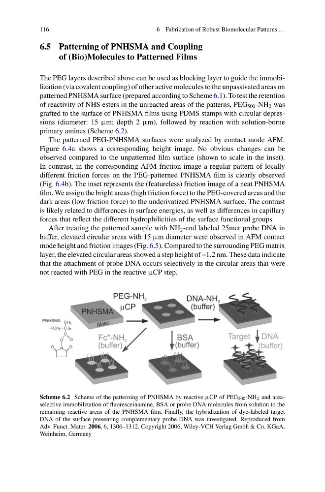
of reactivity of NHS esters in the unreacted areas of the patterns, PEG 500 -NH 2 was
grafted to the surface of PNHSMA films using PDMS stamps with circular depressions (diameter: 15 μm; depth 2 μm), followed by reaction with solution-borne
primary amines (Scheme 6.2).
The patterned PEG-PNHSMA surfaces were analyzed by contact mode AFM.
Figure 6.4a shows a corresponding height image. No obvious changes can be
observed compared to the unpatterned film surface (shown to scale in the inset).
In contrast, in the corresponding AFM friction image a regular pattern of locally
different friction forces on the PEG-patterned PNHSMA film is clearly observed
(Fig. 6.4b). The inset represents the (featureless) friction image of a neat PNHSMA
film. We assign the bright areas (high friction force) to the PEG-covered areas and the
dark areas (low friction force) to the underivatized PNHSMA surface. The contrast
is likely related to differences in surface energies, as well as differences in capillary
forces that reflect the different hydrophilicities of the surface functional groups.
After treating the patterned sample with NH 2 -end labeled 25mer probe DNA in
buffer, elevated circular areas with 15 μm diameter were observed in AFM contact
mode height and friction images (Fig. 6.5). Compared to the surrounding PEG matrix
layer, the elevated circular areas showed a step height of ~1.2 nm. These data indicate
that the attachment of probe DNA occurs selectively in the circular areas that were
not reacted with PEG in the reactive μCP step.
Scheme 6.2 Scheme of the patterning of PNHSMA by reactive μCP of PEG 500 -NH 2 and areaselective immobilization of fluoresceinamine, BSA or probe DNA molecules from solution to the
remaining reactive areas of the PNHSMA film. Finally, the hybridization of dye-labeled target
DNA of the surface presenting complementary probe DNA was investigated. Reproduced from
Adv. Funct. Mater. 2006, 6, 1306–1312. Copyright 2006, Wiley-VCH Verlag Gmbh & Co. KGaA,
Weinheim, Germany
6 Fabrication of Robust Biomolecular Patterns …
6.5 Patterning of PNHSMA and Coupling
of (Bio)Molecules to Patterned Films
The PEG layers described above can be used as blocking layer to guide the immobilization (via covalent coupling) of other active molecules to the unpassivated areas on
patterned PNHSMA surface (prepared according to Scheme 6.1). To test the retention
of reactivity of NHS esters in the unreacted areas of the patterns, PEG 500 -NH 2 was
grafted to the surface of PNHSMA films using PDMS stamps with circular depressions (diameter: 15 μm; depth 2 μm), followed by reaction with solution-borne
primary amines (Scheme 6.2).
The patterned PEG-PNHSMA surfaces were analyzed by contact mode AFM.
Figure 6.4a shows a corresponding height image. No obvious changes can be
observed compared to the unpatterned film surface (shown to scale in the inset).
In contrast, in the corresponding AFM friction image a regular pattern of locally
different friction forces on the PEG-patterned PNHSMA film is clearly observed
(Fig. 6.4b). The inset represents the (featureless) friction image of a neat PNHSMA
film. We assign the bright areas (high friction force) to the PEG-covered areas and the
dark areas (low friction force) to the underivatized PNHSMA surface. The contrast
is likely related to differences in surface energies, as well as differences in capillary
forces that reflect the different hydrophilicities of the surface functional groups.
After treating the patterned sample with NH 2 -end labeled 25mer probe DNA in
buffer, elevated circular areas with 15 μm diameter were observed in AFM contact
mode height and friction images (Fig. 6.5). Compared to the surrounding PEG matrix
layer, the elevated circular areas showed a step height of ~1.2 nm. These data indicate
that the attachment of probe DNA occurs selectively in the circular areas that were
not reacted with PEG in the reactive μCP step.
Scheme 6.2 Scheme of the patterning of PNHSMA by reactive μCP of PEG 500 -NH 2 and areaselective immobilization of fluoresceinamine, BSA or probe DNA molecules from solution to the
remaining reactive areas of the PNHSMA film. Finally, the hybridization of dye-labeled target
DNA of the surface presenting complementary probe DNA was investigated. Reproduced from
Adv. Funct. Mater. 2006, 6, 1306–1312. Copyright 2006, Wiley-VCH Verlag Gmbh & Co. KGaA,
Weinheim, Germany