

3.2 Results
85
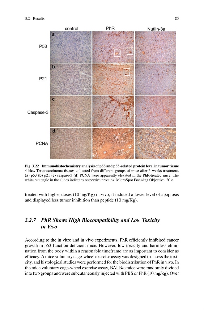
Fig. 3.22 Immunohistochemistry analysis of p53 and p53-related protein level in tumor tissue
slides. Teratocarcinoma tissues collected from different groups of mice after 3 weeks treatment.
(a) p53 (b) p21 (c) caspase-3 (d) PCNA were apparently elevated in the PhR-treated mice. The
white rectangle in the slides indicates respective proteins. MicroSpot Focusing Objective, 20×
treated with higher doses (10 mg/Kg) in vivo, it induced a lower level of apoptosis
and displayed less tumor inhibition than peptide (10 mg/Kg).
3.2.7 PhR Shows High Biocompatibility and Low Toxicity
in Vivo
According to the in vitro and in vivo experiments, PhR efficiently inhibited cancer
growth in p53 function-deficient mice. However, low-toxicity and harmless elimination from the body within a reasonable timeframe are as important to consider as
efficacy. A mice voluntary cage-wheel exercise assay was designed to assess the toxicity, and histological studies were performed for the biodistribution of PhR in vivo. In
the mice voluntary cage-wheel exercise assay, BALB/c mice were randomly divided
into two groups and were subcutaneously injected with PBS or PhR (10 mg/kg). Over
85
Fig. 3.22 Immunohistochemistry analysis of p53 and p53-related protein level in tumor tissue
slides. Teratocarcinoma tissues collected from different groups of mice after 3 weeks treatment.
(a) p53 (b) p21 (c) caspase-3 (d) PCNA were apparently elevated in the PhR-treated mice. The
white rectangle in the slides indicates respective proteins. MicroSpot Focusing Objective, 20×
treated with higher doses (10 mg/Kg) in vivo, it induced a lower level of apoptosis
and displayed less tumor inhibition than peptide (10 mg/Kg).
3.2.7 PhR Shows High Biocompatibility and Low Toxicity
in Vivo
According to the in vitro and in vivo experiments, PhR efficiently inhibited cancer
growth in p53 function-deficient mice. However, low-toxicity and harmless elimination from the body within a reasonable timeframe are as important to consider as
efficacy. A mice voluntary cage-wheel exercise assay was designed to assess the toxicity, and histological studies were performed for the biodistribution of PhR in vivo. In
the mice voluntary cage-wheel exercise assay, BALB/c mice were randomly divided
into two groups and were subcutaneously injected with PBS or PhR (10 mg/kg). Over