

2.2 Results
39
2.2.7 On the Mechanism of Chirality-Induced Helicity
To better understand the effects of the in-tether chiral center on the -helix content, we
carried out simulations on peptides 14 Ac-cyclo-CAAAS 5 -NH 2 (R = H), 10a/10b
(R = CH3), and 2a/2b (R = Ph). Peptides 1a/1b were also simulated (Fig. 2.10a,
b), and results very similar to those of 10a/10b were obtained (Fig. 2.9). Here
we use 10a/10b to discuss because only 10b has the crystal structure. By using
replica-exchange molecular dynamics (REMD) method, converged conformational
sampling was achieved for each peptide. The simulated results are in good agreement
with experimental observations. For peptide 14, the representative structure is nonhelical, with four out of five residues in PII conformation (Fig. 2.9a). Indeed, from
its Ramachandran (φ, ψ) plot, PII is more favored than the and β conformations.
This can be understood by the recent findings that PII conformation is intrinsically
preferred for short unfolded peptides in aqueous solution. The representative structure of peptide 10a with (S)-chirality in the tether is very similar to that of peptide 14,
with the added methyl group point outward without any steric interference (Fig. 2.9b).
Therefore, as in the case of 14, the non-helical structure of 10a is also stable. The
peptide 2a with R = Ph also has the similar situation (Fig. 2.9c).
However, this non-helical structure will be significantly destabilized when a
methyl substitution is placed at (R)-configuration, due to severe steric clash with
the backbone O atom on the third residue (Fig. 2.9b). To relieve this clash, the tether
chain will rotate to make the methyl group away from this O atom, as the Cluster 2 in
Fig. 2.9c. However, the energy may increase due to the conformational change in the
tether. Also, the methyl group becomes in contact with another backbone O atom,
probably interfere with its solvation by water. Thus, the most populated structure
cluster for peptides 10b is α-helical, in which the R group is quite comfortable, as
the representative structure of the Cluster 1 in Figure S5c. This simulated structure is
almost identical to the crystal structure, with RMSD of backbone and Cβ only 0.3Å
(Fig. 2.9c).
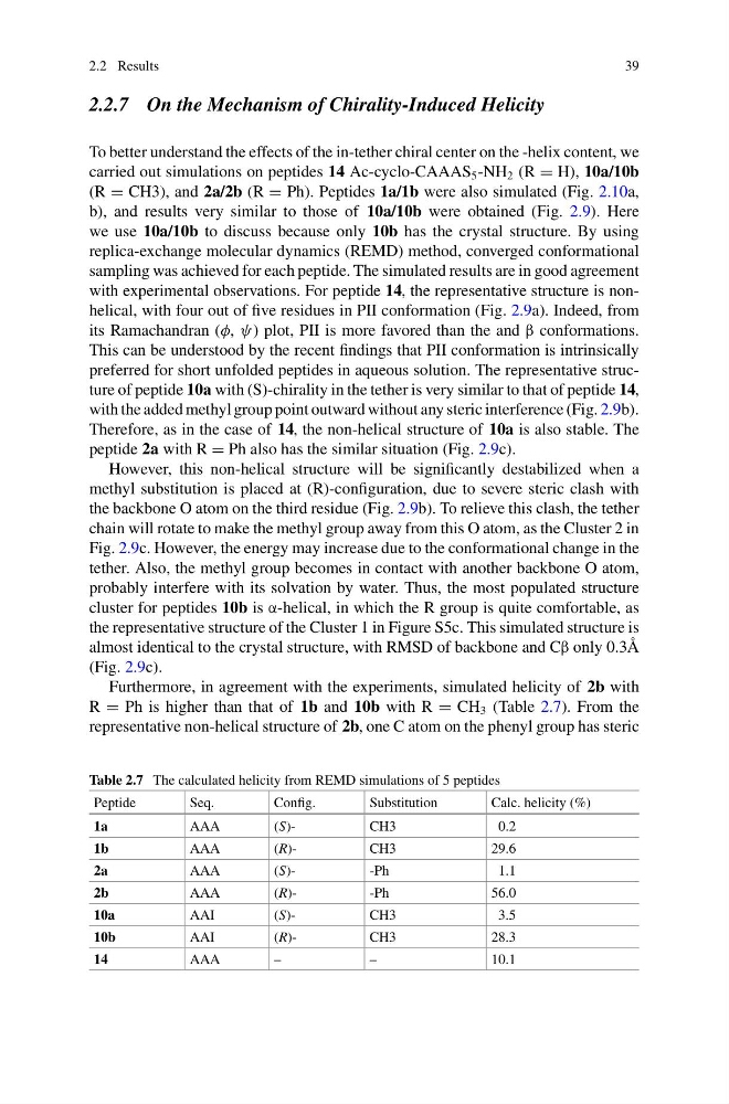
Furthermore, in agreement with the experiments, simulated helicity of 2b with
R = Ph is higher than that of 1b and 10b with R = CH 3 (Table 2.7). From the
representative non-helical structure of 2b, one C atom on the phenyl group has steric
Table 2.7 The calculated helicity from REMD simulations of 5 peptides
Peptide
Seq.
Config.
Substitution
Calc. helicity (%)
1a
AAA
(S)CH3
0.2
1b
AAA
(R)CH3
29.6
2a
AAA
(S)-Ph
1.1
2b
AAA
(R)-Ph
56.0
10a
AAI
(S)CH3
3.5
10b
AAI
(R)CH3
28.3
14
AAA
–
–
10.1
39
2.2.7 On the Mechanism of Chirality-Induced Helicity
To better understand the effects of the in-tether chiral center on the -helix content, we
carried out simulations on peptides 14 Ac-cyclo-CAAAS 5 -NH 2 (R = H), 10a/10b
(R = CH3), and 2a/2b (R = Ph). Peptides 1a/1b were also simulated (Fig. 2.10a,
b), and results very similar to those of 10a/10b were obtained (Fig. 2.9). Here
we use 10a/10b to discuss because only 10b has the crystal structure. By using
replica-exchange molecular dynamics (REMD) method, converged conformational
sampling was achieved for each peptide. The simulated results are in good agreement
with experimental observations. For peptide 14, the representative structure is nonhelical, with four out of five residues in PII conformation (Fig. 2.9a). Indeed, from
its Ramachandran (φ, ψ) plot, PII is more favored than the and β conformations.
This can be understood by the recent findings that PII conformation is intrinsically
preferred for short unfolded peptides in aqueous solution. The representative structure of peptide 10a with (S)-chirality in the tether is very similar to that of peptide 14,
with the added methyl group point outward without any steric interference (Fig. 2.9b).
Therefore, as in the case of 14, the non-helical structure of 10a is also stable. The
peptide 2a with R = Ph also has the similar situation (Fig. 2.9c).
However, this non-helical structure will be significantly destabilized when a
methyl substitution is placed at (R)-configuration, due to severe steric clash with
the backbone O atom on the third residue (Fig. 2.9b). To relieve this clash, the tether
chain will rotate to make the methyl group away from this O atom, as the Cluster 2 in
Fig. 2.9c. However, the energy may increase due to the conformational change in the
tether. Also, the methyl group becomes in contact with another backbone O atom,
probably interfere with its solvation by water. Thus, the most populated structure
cluster for peptides 10b is α-helical, in which the R group is quite comfortable, as
the representative structure of the Cluster 1 in Figure S5c. This simulated structure is
almost identical to the crystal structure, with RMSD of backbone and Cβ only 0.3Å
(Fig. 2.9c).
Furthermore, in agreement with the experiments, simulated helicity of 2b with
R = Ph is higher than that of 1b and 10b with R = CH 3 (Table 2.7). From the
representative non-helical structure of 2b, one C atom on the phenyl group has steric
Table 2.7 The calculated helicity from REMD simulations of 5 peptides
Peptide
Seq.
Config.
Substitution
Calc. helicity (%)
1a
AAA
(S)CH3
0.2
1b
AAA
(R)CH3
29.6
2a
AAA
(S)-Ph
1.1
2b
AAA
(R)-Ph
56.0
10a
AAI
(S)CH3
3.5
10b
AAI
(R)CH3
28.3
14
AAA
–
–
10.1