

2.2 Results
37
Table 2.5 Hydrogen bonds parameters of peptide crystal. The distance of H…O atoms at the i, i
+ 4 positions is less than 2.8 Å, which means forming strong intramolecular hydrogen bond
Parameters of the hydrogen bonds (Å,°)
Donor
Acceptor
N…O
H…O
N-H…O
C-O…H
N-NH2
O4
2.84
2.03
152
143
N1
O5
2.88
2.02
163
156
N2
O-Ac
3.05
2.22
158
156
Table 2.6 The dihedral
angles of solved crystal. 4 of
5 pairs of parameters except
the S 5 closed to standard
alpha helix means 10b
formed helix in H 2 O
Residue
ϕ
ψ
ω
S 5 1
−92
−22
−166
I2
−68
−53
−180
A4
−62
−38
−177
A4
−63
−42
−179
C5
−57
−42
−130
a
b
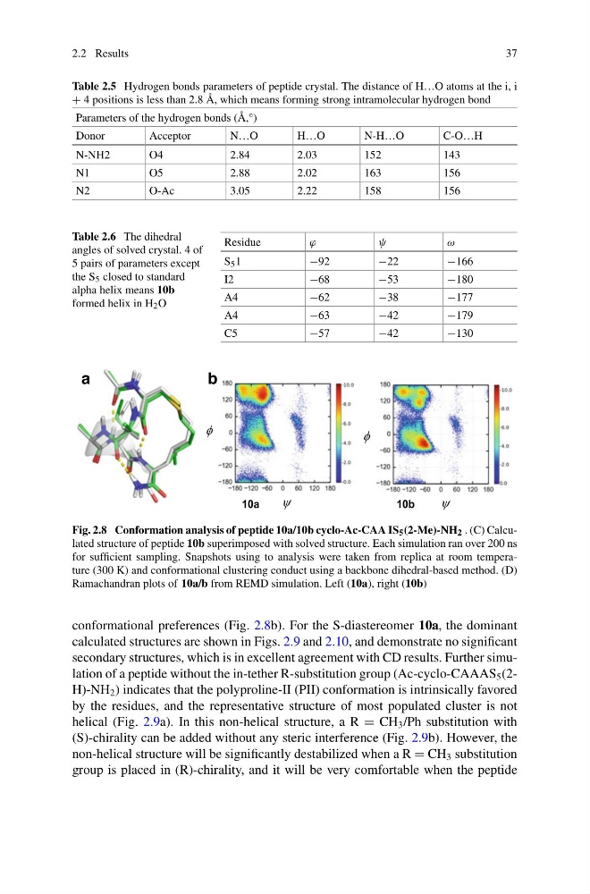
Fig. 2.8 Conformation analysis of peptide 10a/10b cyclo-Ac-CAA IS 5 (2-Me)-NH 2 . (C) Calculated structure of peptide 10b superimposed with solved structure. Each simulation ran over 200 ns
for sufficient sampling. Snapshots using to analysis were taken from replica at room temperature (300 K) and conformational clustering conduct using a backbone dihedral-based method. (D)
Ramachandran plots of 10a/b from REMD simulation. Left (10a), right (10b)
conformational preferences (Fig. 2.8b). For the S-diastereomer 10a, the dominant
calculated structures are shown in Figs. 2.9 and 2.10, and demonstrate no significant
secondary structures, which is in excellent agreement with CD results. Further simulation of a peptide without the in-tether R-substitution group (Ac-cyclo-CAAAS 5 (2H)-NH 2 ) indicates that the polyproline-II (PII) conformation is intrinsically favored
by the residues, and the representative structure of most populated cluster is not
helical (Fig. 2.9a). In this non-helical structure, a R = CH 3 /Ph substitution with
(S)-chirality can be added without any steric interference (Fig. 2.9b). However, the
non-helical structure will be significantly destabilized when a R = CH 3 substitution
group is placed in (R)-chirality, and it will be very comfortable when the peptide
37
Table 2.5 Hydrogen bonds parameters of peptide crystal. The distance of H…O atoms at the i, i
+ 4 positions is less than 2.8 Å, which means forming strong intramolecular hydrogen bond
Parameters of the hydrogen bonds (Å,°)
Donor
Acceptor
N…O
H…O
N-H…O
C-O…H
N-NH2
O4
2.84
2.03
152
143
N1
O5
2.88
2.02
163
156
N2
O-Ac
3.05
2.22
158
156
Table 2.6 The dihedral
angles of solved crystal. 4 of
5 pairs of parameters except
the S 5 closed to standard
alpha helix means 10b
formed helix in H 2 O
Residue
ϕ
ψ
ω
S 5 1
−92
−22
−166
I2
−68
−53
−180
A4
−62
−38
−177
A4
−63
−42
−179
C5
−57
−42
−130
a
b
Fig. 2.8 Conformation analysis of peptide 10a/10b cyclo-Ac-CAA IS 5 (2-Me)-NH 2 . (C) Calculated structure of peptide 10b superimposed with solved structure. Each simulation ran over 200 ns
for sufficient sampling. Snapshots using to analysis were taken from replica at room temperature (300 K) and conformational clustering conduct using a backbone dihedral-based method. (D)
Ramachandran plots of 10a/b from REMD simulation. Left (10a), right (10b)
conformational preferences (Fig. 2.8b). For the S-diastereomer 10a, the dominant
calculated structures are shown in Figs. 2.9 and 2.10, and demonstrate no significant
secondary structures, which is in excellent agreement with CD results. Further simulation of a peptide without the in-tether R-substitution group (Ac-cyclo-CAAAS 5 (2H)-NH 2 ) indicates that the polyproline-II (PII) conformation is intrinsically favored
by the residues, and the representative structure of most populated cluster is not
helical (Fig. 2.9a). In this non-helical structure, a R = CH 3 /Ph substitution with
(S)-chirality can be added without any steric interference (Fig. 2.9b). However, the
non-helical structure will be significantly destabilized when a R = CH 3 substitution
group is placed in (R)-chirality, and it will be very comfortable when the peptide