

74
involved the yolk sac, the heart, the nervous system, the head, structures derived
from the branchial arches, and the forelimb (Jung et al. 2015).
While the cited works demonstrated the usefulness and the richness of information of the whole embryo culture, to our knowledge, there are no other works implementing it to study nanoparticles.
3.5.3 The Multipotent Embryonic Stem Cells
The embryonic stem cells represent a versatile system to develop different strategies
to study in vitro the developmental toxicity. Derived from the inner cells of early
blastocyst of different species, they form in vitro a cohesive monolayer that retains
the multipotency of the original cells, in the presence of feeder cells and factors that
contrast the differentiation. In their absence, the embryonic stem cells start to
differentiate and grow as three-dimensional clusters, the embryoid bodies, in which
the differentiation spontaneously begins. The embryonic stem cell test approved by
the European Centre for the Validation of Alternative Methods included three
endpoints, the inhibition of 50% of growth of D3 stem cells line and of mouse
embryo fibroblasts (IC50 D3 or other ESCs lines, and IC50 3 T3, respectively), and
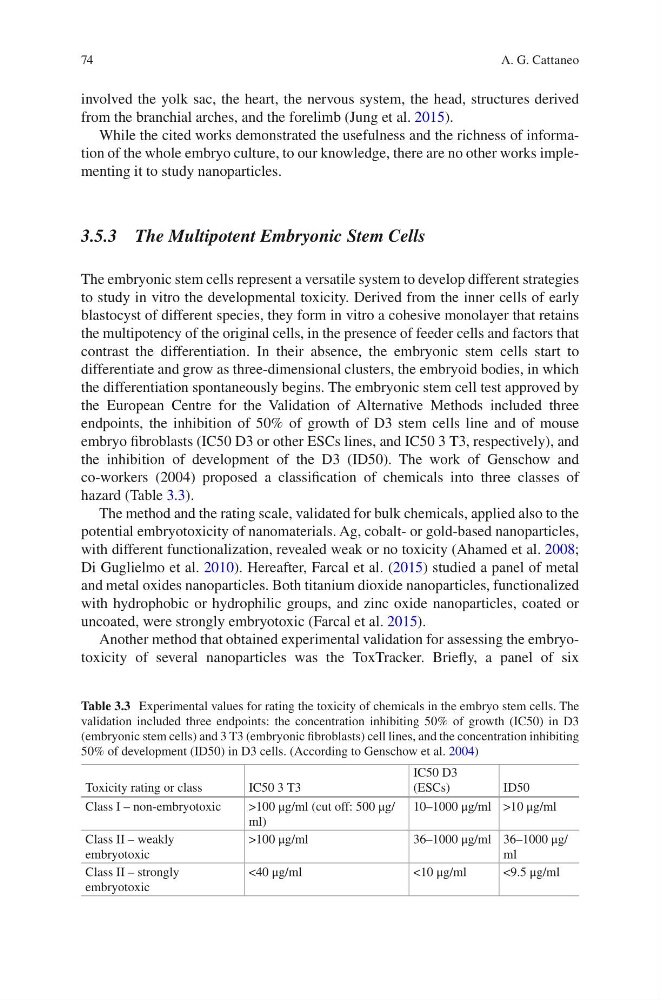
the inhibition of development of the D3 (ID50). The work of Genschow and
co-workers (2004) proposed a classification of chemicals into three classes of
hazard (Table 3.3).
The method and the rating scale, validated for bulk chemicals, applied also to the
potential embryotoxicity of nanomaterials. Ag, cobalt- or gold-based nanoparticles,
with different functionalization, revealed weak or no toxicity (Ahamed et al. 2008;
Di Guglielmo et al. 2010). Hereafter, Farcal et al. (2015) studied a panel of metal
and metal oxides nanoparticles. Both titanium dioxide nanoparticles, functionalized
with hydrophobic or hydrophilic groups, and zinc oxide nanoparticles, coated or
uncoated, were strongly embryotoxic (Farcal et al. 2015).
Another method that obtained experimental validation for assessing the embryotoxicity of several nanoparticles was the ToxTracker. Briefly, a panel of six
Table 3.3 Experimental values for rating the toxicity of chemicals in the embryo stem cells. The
validation included three endpoints: the concentration inhibiting 50% of growth (IC50) in D3
(embryonic stem cells) and 3 T3 (embryonic fibroblasts) cell lines, and the concentration inhibiting
50% of development (ID50) in D3 cells. (According to Genschow et al. 2004)
Toxicity rating or class
IC50 3 T3
IC50 D3
(ESCs)
ID50
Class I – non-embryotoxic
>100 μg/ml (cut off: 500 μg/
ml)
10–1000 μg/ml >10 μg/ml
Class II – weakly
embryotoxic
>100 μg/ml
36–1000 μg/ml 36–1000 μg/
ml
Class II – strongly
embryotoxic
<40 μg/ml
<10 μg/ml
<9.5 μg/ml
A. G. Cattaneo
involved the yolk sac, the heart, the nervous system, the head, structures derived
from the branchial arches, and the forelimb (Jung et al. 2015).
While the cited works demonstrated the usefulness and the richness of information of the whole embryo culture, to our knowledge, there are no other works implementing it to study nanoparticles.
3.5.3 The Multipotent Embryonic Stem Cells
The embryonic stem cells represent a versatile system to develop different strategies
to study in vitro the developmental toxicity. Derived from the inner cells of early
blastocyst of different species, they form in vitro a cohesive monolayer that retains
the multipotency of the original cells, in the presence of feeder cells and factors that
contrast the differentiation. In their absence, the embryonic stem cells start to
differentiate and grow as three-dimensional clusters, the embryoid bodies, in which
the differentiation spontaneously begins. The embryonic stem cell test approved by
the European Centre for the Validation of Alternative Methods included three
endpoints, the inhibition of 50% of growth of D3 stem cells line and of mouse
embryo fibroblasts (IC50 D3 or other ESCs lines, and IC50 3 T3, respectively), and
the inhibition of development of the D3 (ID50). The work of Genschow and
co-workers (2004) proposed a classification of chemicals into three classes of
hazard (Table 3.3).
The method and the rating scale, validated for bulk chemicals, applied also to the
potential embryotoxicity of nanomaterials. Ag, cobalt- or gold-based nanoparticles,
with different functionalization, revealed weak or no toxicity (Ahamed et al. 2008;
Di Guglielmo et al. 2010). Hereafter, Farcal et al. (2015) studied a panel of metal
and metal oxides nanoparticles. Both titanium dioxide nanoparticles, functionalized
with hydrophobic or hydrophilic groups, and zinc oxide nanoparticles, coated or
uncoated, were strongly embryotoxic (Farcal et al. 2015).
Another method that obtained experimental validation for assessing the embryotoxicity of several nanoparticles was the ToxTracker. Briefly, a panel of six
Table 3.3 Experimental values for rating the toxicity of chemicals in the embryo stem cells. The
validation included three endpoints: the concentration inhibiting 50% of growth (IC50) in D3
(embryonic stem cells) and 3 T3 (embryonic fibroblasts) cell lines, and the concentration inhibiting
50% of development (ID50) in D3 cells. (According to Genschow et al. 2004)
Toxicity rating or class
IC50 3 T3
IC50 D3
(ESCs)
ID50
Class I – non-embryotoxic
>100 μg/ml (cut off: 500 μg/
ml)
10–1000 μg/ml >10 μg/ml
Class II – weakly
embryotoxic
>100 μg/ml
36–1000 μg/ml 36–1000 μg/
ml
Class II – strongly
embryotoxic
<40 μg/ml
<10 μg/ml
<9.5 μg/ml
A. G. Cattaneo