

72
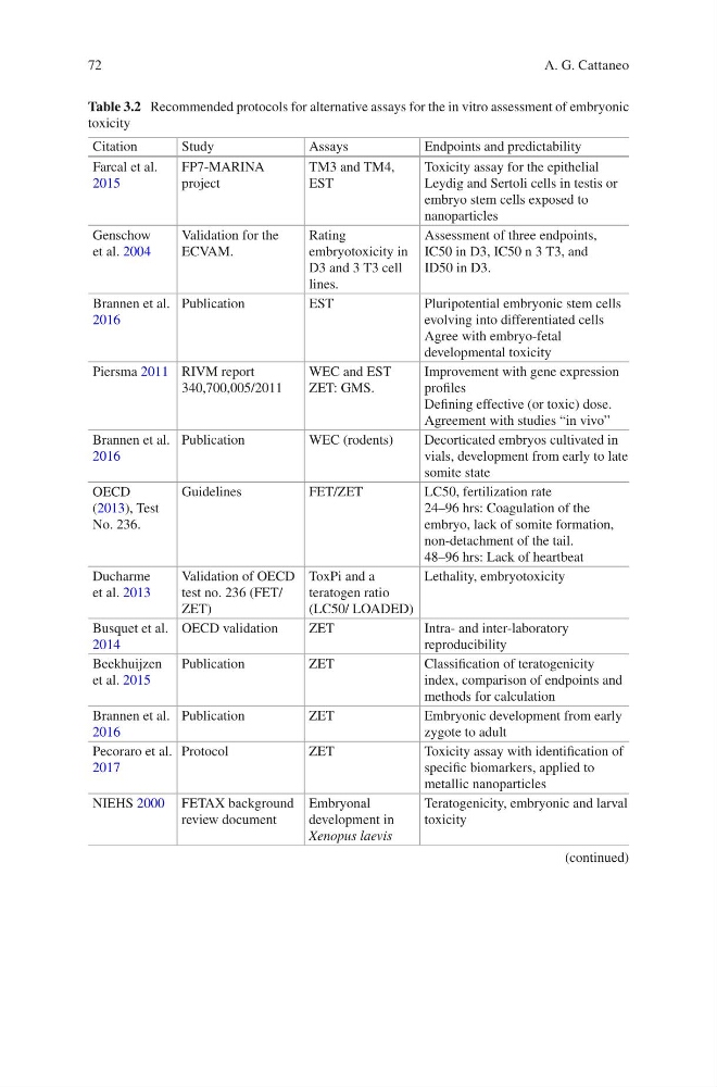
Table 3.2 Recommended protocols for alternative assays for the in vitro assessment of embryonic
toxicity
Citation
Study
Assays
Endpoints and predictability
Farcal et al.
2015
FP7-MARINA
project
TM3 and TM4,
EST
Toxicity assay for the epithelial
Leydig and Sertoli cells in testis or
embryo stem cells exposed to
nanoparticles
Genschow
et al. 2004
Validation for the
ECVAM.
Rating
embryotoxicity in
D3 and 3 T3 cell
lines.
Assessment of three endpoints,
IC50 in D3, IC50 n 3 T3, and
ID50 in D3.
Brannen et al.
2016
Publication
EST
Pluripotential embryonic stem cells
evolving into differentiated cells
Agree with embryo-fetal
developmental toxicity
Piersma 2011 RIVM report
340,700,005/2011
WEC and EST
ZET: GMS.
Improvement with gene expression
profiles
Defining effective (or toxic) dose.
Agreement with studies “in vivo”
Brannen et al.
2016
Publication
WEC (rodents)
Decorticated embryos cultivated in
vials, development from early to late
somite state
OECD
(2013), Test
No. 236.
Guidelines
FET/ZET
LC50, fertilization rate
24–96 hrs: Coagulation of the
embryo, lack of somite formation,
non-detachment of the tail.
48–96 hrs: Lack of heartbeat
Ducharme
et al. 2013
Validation of OECD
test no. 236 (FET/
ZET)
ToxPi and a
teratogen ratio
(LC50/ LOADED)
Lethality, embryotoxicity
Busquet et al.
2014
OECD validation
ZET
Intra- and inter-laboratory
reproducibility
Beekhuijzen
et al. 2015
Publication
ZET
Classification of teratogenicity
index, comparison of endpoints and
methods for calculation
Brannen et al.
2016
Publication
ZET
Embryonic development from early
zygote to adult
Pecoraro et al.
2017
Protocol
ZET
Toxicity assay with identification of
specific biomarkers, applied to
metallic nanoparticles
NIEHS 2000 FETAX background
review document
Embryonal
development in
Xenopus laevis
Teratogenicity, embryonic and larval
toxicity
(continued)
A. G. Cattaneo
Table 3.2 Recommended protocols for alternative assays for the in vitro assessment of embryonic
toxicity
Citation
Study
Assays
Endpoints and predictability
Farcal et al.
2015
FP7-MARINA
project
TM3 and TM4,
EST
Toxicity assay for the epithelial
Leydig and Sertoli cells in testis or
embryo stem cells exposed to
nanoparticles
Genschow
et al. 2004
Validation for the
ECVAM.
Rating
embryotoxicity in
D3 and 3 T3 cell
lines.
Assessment of three endpoints,
IC50 in D3, IC50 n 3 T3, and
ID50 in D3.
Brannen et al.
2016
Publication
EST
Pluripotential embryonic stem cells
evolving into differentiated cells
Agree with embryo-fetal
developmental toxicity
Piersma 2011 RIVM report
340,700,005/2011
WEC and EST
ZET: GMS.
Improvement with gene expression
profiles
Defining effective (or toxic) dose.
Agreement with studies “in vivo”
Brannen et al.
2016
Publication
WEC (rodents)
Decorticated embryos cultivated in
vials, development from early to late
somite state
OECD
(2013), Test
No. 236.
Guidelines
FET/ZET
LC50, fertilization rate
24–96 hrs: Coagulation of the
embryo, lack of somite formation,
non-detachment of the tail.
48–96 hrs: Lack of heartbeat
Ducharme
et al. 2013
Validation of OECD
test no. 236 (FET/
ZET)
ToxPi and a
teratogen ratio
(LC50/ LOADED)
Lethality, embryotoxicity
Busquet et al.
2014
OECD validation
ZET
Intra- and inter-laboratory
reproducibility
Beekhuijzen
et al. 2015
Publication
ZET
Classification of teratogenicity
index, comparison of endpoints and
methods for calculation
Brannen et al.
2016
Publication
ZET
Embryonic development from early
zygote to adult
Pecoraro et al.
2017
Protocol
ZET
Toxicity assay with identification of
specific biomarkers, applied to
metallic nanoparticles
NIEHS 2000 FETAX background
review document
Embryonal
development in
Xenopus laevis
Teratogenicity, embryonic and larval
toxicity
(continued)
A. G. Cattaneo