

66
particles, and therefore protects the zygote without impeding the necessary
exchanges with the mother.
The placenta develops in two times, beginning with a choriovitelline placenta,
replaced by the well-vascularized chorioallantoic placenta, and when the zygote
needs higher oxygen supply. Species with short pregnancy, such as mice and rat,
develop the chorioallantoic placenta for a very short time. This organ is substantially
different in humans and in the most popular model species and recommended
models for chemical toxicity in vivo, such as rodents, rabbits, and minipig. The
histological features, known from decades, differ between families and are conserved
inside the orders of mammals (Enders 1965, 2009; Enders and Blankenship 1999).
The histology and permeability of the placentas in livestock have been recently
reviewed and compared (Furukawa et al. 2014). Several details are reported in
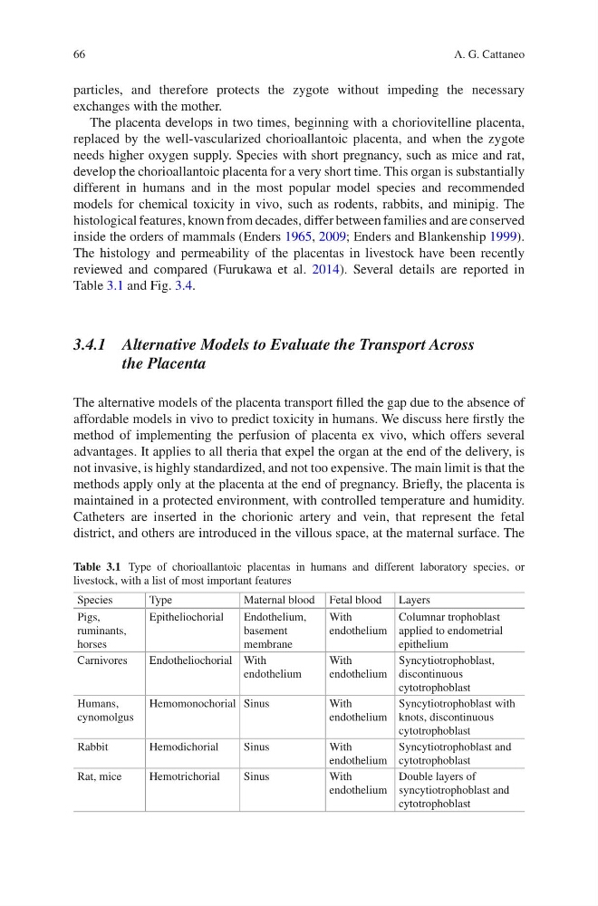
Table 3.1 and Fig. 3.4.
3.4.1 Alternative Models to Evaluate the Transport Across
the Placenta
The alternative models of the placenta transport filled the gap due to the absence of
affordable models in vivo to predict toxicity in humans. We discuss here firstly the
method of implementing the perfusion of placenta ex vivo, which offers several
advantages. It applies to all theria that expel the organ at the end of the delivery, is
not invasive, is highly standardized, and not too expensive. The main limit is that the
methods apply only at the placenta at the end of pregnancy. Briefly, the placenta is
maintained in a protected environment, with controlled temperature and humidity.
Catheters are inserted in the chorionic artery and vein, that represent the fetal
district, and others are introduced in the villous space, at the maternal surface. The
Table 3.1 Type of chorioallantoic placentas in humans and different laboratory species, or
livestock, with a list of most important features
Species
Type
Maternal blood Fetal blood Layers
Pigs,
ruminants,
horses
Epitheliochorial
Endothelium,
basement
membrane
With
endothelium
Columnar trophoblast
applied to endometrial
epithelium
Carnivores
Endotheliochorial With
endothelium
With
endothelium
Syncytiotrophoblast,
discontinuous
cytotrophoblast
Humans,
cynomolgus
Hemomonochorial Sinus
With
endothelium
Syncytiotrophoblast with
knots, discontinuous
cytotrophoblast
Rabbit
Hemodichorial
Sinus
With
endothelium
Syncytiotrophoblast and
cytotrophoblast
Rat, mice
Hemotrichorial
Sinus
With
endothelium
Double layers of
syncytiotrophoblast and
cytotrophoblast
A. G. Cattaneo
particles, and therefore protects the zygote without impeding the necessary
exchanges with the mother.
The placenta develops in two times, beginning with a choriovitelline placenta,
replaced by the well-vascularized chorioallantoic placenta, and when the zygote
needs higher oxygen supply. Species with short pregnancy, such as mice and rat,
develop the chorioallantoic placenta for a very short time. This organ is substantially
different in humans and in the most popular model species and recommended
models for chemical toxicity in vivo, such as rodents, rabbits, and minipig. The
histological features, known from decades, differ between families and are conserved
inside the orders of mammals (Enders 1965, 2009; Enders and Blankenship 1999).
The histology and permeability of the placentas in livestock have been recently
reviewed and compared (Furukawa et al. 2014). Several details are reported in
Table 3.1 and Fig. 3.4.
3.4.1 Alternative Models to Evaluate the Transport Across
the Placenta
The alternative models of the placenta transport filled the gap due to the absence of
affordable models in vivo to predict toxicity in humans. We discuss here firstly the
method of implementing the perfusion of placenta ex vivo, which offers several
advantages. It applies to all theria that expel the organ at the end of the delivery, is
not invasive, is highly standardized, and not too expensive. The main limit is that the
methods apply only at the placenta at the end of pregnancy. Briefly, the placenta is
maintained in a protected environment, with controlled temperature and humidity.
Catheters are inserted in the chorionic artery and vein, that represent the fetal
district, and others are introduced in the villous space, at the maternal surface. The
Table 3.1 Type of chorioallantoic placentas in humans and different laboratory species, or
livestock, with a list of most important features
Species
Type
Maternal blood Fetal blood Layers
Pigs,
ruminants,
horses
Epitheliochorial
Endothelium,
basement
membrane
With
endothelium
Columnar trophoblast
applied to endometrial
epithelium
Carnivores
Endotheliochorial With
endothelium
With
endothelium
Syncytiotrophoblast,
discontinuous
cytotrophoblast
Humans,
cynomolgus
Hemomonochorial Sinus
With
endothelium
Syncytiotrophoblast with
knots, discontinuous
cytotrophoblast
Rabbit
Hemodichorial
Sinus
With
endothelium
Syncytiotrophoblast and
cytotrophoblast
Rat, mice
Hemotrichorial
Sinus
With
endothelium
Double layers of
syncytiotrophoblast and
cytotrophoblast
A. G. Cattaneo