

2.2 Discharges of Current Large Tokamaks
21
TFTR
(a)
(c)
(b)
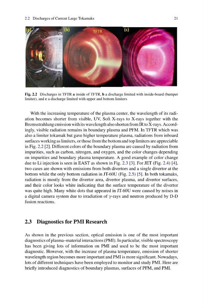
Fig. 2.2 Discharges in TFTR a inside of TFTR, b a discharge limited with inside-board (bumper
limiter), and c a discharge limited with upper and bottom limiters
With the increasing temperature of the plasma center, the wavelength of its radiation becomes shorter from visible, UV, Soft X-rays to X-rays together with the
Bremsstrahlung emission with its wavelength also shorten from IR to X-rays. Accordingly, visible radiation remains in boundary plasma and PFM. In TFTR which was
also a limiter tokamak but gave higher temperature plasma, radiations from inboard
surfaces working as limiters, or those from the bottom and top limiters are appreciable
in Fig. 2.2 [2]. Different colors of the boundary plasma are caused by radiation from
impurities, such as carbon, nitrogen, and oxygen, and the color changes depending
on impurities and boundary plasma temperature. A good example of color change
due to Li injection is seen in EAST as shown in Fig. 2.3 [3]. For JET (Fig. 2.4) [4],
two cases are shown with emissions from both divertors and a single divertor at the
bottom while the only bottom radiation in JT-60U (Fig. 2.5) [5]. In both tokamaks,
radiation is mostly from the divertor area, divertor plasma, and divertor surfaces,
and their color looks white indicating that the surface temperature of the divertor
was quite high. Many white dots that appeared in JT-60U were caused by noises in
a digital camera system due to irradiation of γ-rays and neutron produced by D-D
fusion reactions.
2.3 Diagnostics for PMI Research
As shown in the previous section, optical emission is one of the most important
diagnostics of plasma–material interactions (PMI). In particular, visible spectroscopy
has been giving lots of information on PMI and used to be the most important
diagnostic. However, with the increase of plasma temperature, emission of shorter
wavelength region becomes more important and PMI is more significant. Nowadays,
lots of different techniques have been employed to monitor and study PMI. Here are
briefly introduced diagnostics of boundary plasmas, surfaces of PFM, and PMI.
21
TFTR
(a)
(c)
(b)
Fig. 2.2 Discharges in TFTR a inside of TFTR, b a discharge limited with inside-board (bumper
limiter), and c a discharge limited with upper and bottom limiters
With the increasing temperature of the plasma center, the wavelength of its radiation becomes shorter from visible, UV, Soft X-rays to X-rays together with the
Bremsstrahlung emission with its wavelength also shorten from IR to X-rays. Accordingly, visible radiation remains in boundary plasma and PFM. In TFTR which was
also a limiter tokamak but gave higher temperature plasma, radiations from inboard
surfaces working as limiters, or those from the bottom and top limiters are appreciable
in Fig. 2.2 [2]. Different colors of the boundary plasma are caused by radiation from
impurities, such as carbon, nitrogen, and oxygen, and the color changes depending
on impurities and boundary plasma temperature. A good example of color change
due to Li injection is seen in EAST as shown in Fig. 2.3 [3]. For JET (Fig. 2.4) [4],
two cases are shown with emissions from both divertors and a single divertor at the
bottom while the only bottom radiation in JT-60U (Fig. 2.5) [5]. In both tokamaks,
radiation is mostly from the divertor area, divertor plasma, and divertor surfaces,
and their color looks white indicating that the surface temperature of the divertor
was quite high. Many white dots that appeared in JT-60U were caused by noises in
a digital camera system due to irradiation of γ-rays and neutron produced by D-D
fusion reactions.
2.3 Diagnostics for PMI Research
As shown in the previous section, optical emission is one of the most important
diagnostics of plasma–material interactions (PMI). In particular, visible spectroscopy
has been giving lots of information on PMI and used to be the most important
diagnostic. However, with the increase of plasma temperature, emission of shorter
wavelength region becomes more important and PMI is more significant. Nowadays,
lots of different techniques have been employed to monitor and study PMI. Here are
briefly introduced diagnostics of boundary plasmas, surfaces of PFM, and PMI.