

2.3 Crucial Importance of Hydration Entropy …
9
Solute 2
Solute 1
Solute 2
System (I)
System (III)
Solute 1
Solute 2
System (II)
Solute 1
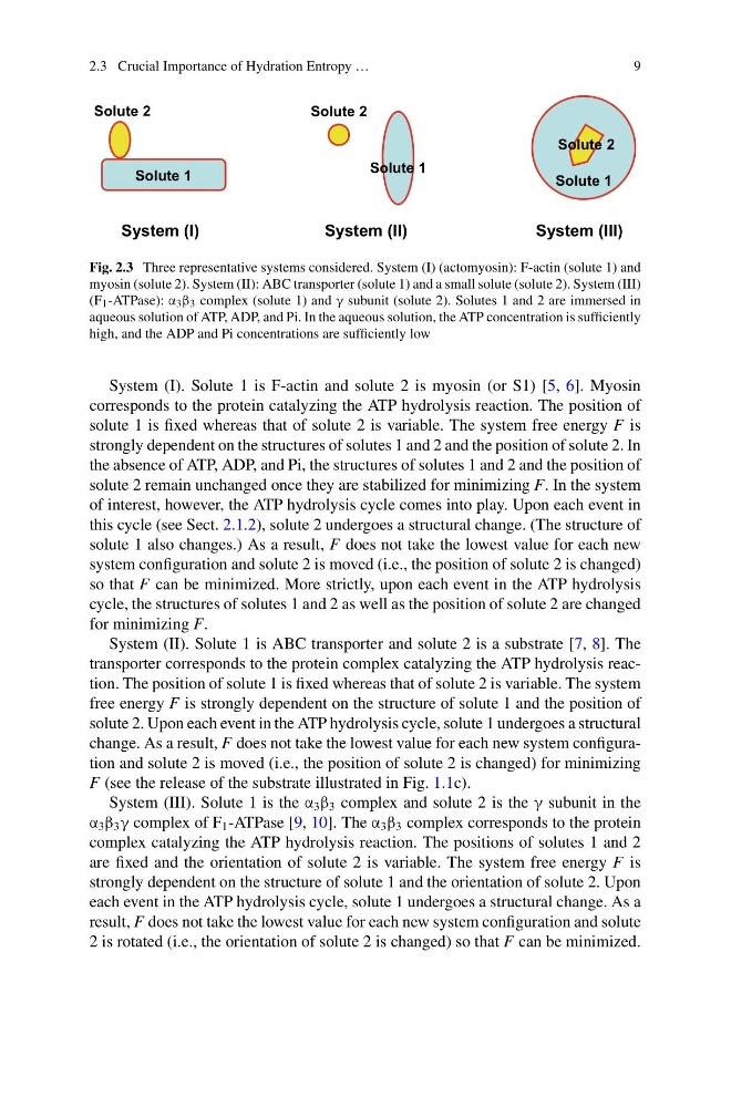
Fig. 2.3 Three representative systems considered. System (I) (actomyosin): F-actin (solute 1) and
myosin (solute 2). System (II): ABC transporter (solute 1) and a small solute (solute 2). System (III)
(F 1 -ATPase): α 3 β 3 complex (solute 1) and γ subunit (solute 2). Solutes 1 and 2 are immersed in
aqueous solution of ATP, ADP, and Pi. In the aqueous solution, the ATP concentration is sufficiently
high, and the ADP and Pi concentrations are sufficiently low
System (I). Solute 1 is F-actin and solute 2 is myosin (or S1) [5, 6]. Myosin
corresponds to the protein catalyzing the ATP hydrolysis reaction. The position of
solute 1 is fixed whereas that of solute 2 is variable. The system free energy F is
strongly dependent on the structures of solutes 1 and 2 and the position of solute 2. In
the absence of ATP, ADP, and Pi, the structures of solutes 1 and 2 and the position of
solute 2 remain unchanged once they are stabilized for minimizing F. In the system
of interest, however, the ATP hydrolysis cycle comes into play. Upon each event in
this cycle (see Sect. 2.1.2), solute 2 undergoes a structural change. (The structure of
solute 1 also changes.) As a result, F does not take the lowest value for each new
system configuration and solute 2 is moved (i.e., the position of solute 2 is changed)
so that F can be minimized. More strictly, upon each event in the ATP hydrolysis
cycle, the structures of solutes 1 and 2 as well as the position of solute 2 are changed
for minimizing F.
System (II). Solute 1 is ABC transporter and solute 2 is a substrate [7, 8]. The
transporter corresponds to the protein complex catalyzing the ATP hydrolysis reaction. The position of solute 1 is fixed whereas that of solute 2 is variable. The system
free energy F is strongly dependent on the structure of solute 1 and the position of
solute 2. Upon each event in the ATP hydrolysis cycle, solute 1 undergoes a structural
change. As a result, F does not take the lowest value for each new system configuration and solute 2 is moved (i.e., the position of solute 2 is changed) for minimizing
F (see the release of the substrate illustrated in Fig. 1.1c).
System (III). Solute 1 is the α 3 β 3 complex and solute 2 is the γ subunit in the
α 3 β 3 γ complex of F 1 -ATPase [9, 10]. The α 3 β 3 complex corresponds to the protein
complex catalyzing the ATP hydrolysis reaction. The positions of solutes 1 and 2
are fixed and the orientation of solute 2 is variable. The system free energy F is
strongly dependent on the structure of solute 1 and the orientation of solute 2. Upon
each event in the ATP hydrolysis cycle, solute 1 undergoes a structural change. As a
result, F does not take the lowest value for each new system configuration and solute
2 is rotated (i.e., the orientation of solute 2 is changed) so that F can be minimized.
9
Solute 2
Solute 1
Solute 2
System (I)
System (III)
Solute 1
Solute 2
System (II)
Solute 1
Fig. 2.3 Three representative systems considered. System (I) (actomyosin): F-actin (solute 1) and
myosin (solute 2). System (II): ABC transporter (solute 1) and a small solute (solute 2). System (III)
(F 1 -ATPase): α 3 β 3 complex (solute 1) and γ subunit (solute 2). Solutes 1 and 2 are immersed in
aqueous solution of ATP, ADP, and Pi. In the aqueous solution, the ATP concentration is sufficiently
high, and the ADP and Pi concentrations are sufficiently low
System (I). Solute 1 is F-actin and solute 2 is myosin (or S1) [5, 6]. Myosin
corresponds to the protein catalyzing the ATP hydrolysis reaction. The position of
solute 1 is fixed whereas that of solute 2 is variable. The system free energy F is
strongly dependent on the structures of solutes 1 and 2 and the position of solute 2. In
the absence of ATP, ADP, and Pi, the structures of solutes 1 and 2 and the position of
solute 2 remain unchanged once they are stabilized for minimizing F. In the system
of interest, however, the ATP hydrolysis cycle comes into play. Upon each event in
this cycle (see Sect. 2.1.2), solute 2 undergoes a structural change. (The structure of
solute 1 also changes.) As a result, F does not take the lowest value for each new
system configuration and solute 2 is moved (i.e., the position of solute 2 is changed)
so that F can be minimized. More strictly, upon each event in the ATP hydrolysis
cycle, the structures of solutes 1 and 2 as well as the position of solute 2 are changed
for minimizing F.
System (II). Solute 1 is ABC transporter and solute 2 is a substrate [7, 8]. The
transporter corresponds to the protein complex catalyzing the ATP hydrolysis reaction. The position of solute 1 is fixed whereas that of solute 2 is variable. The system
free energy F is strongly dependent on the structure of solute 1 and the position of
solute 2. Upon each event in the ATP hydrolysis cycle, solute 1 undergoes a structural
change. As a result, F does not take the lowest value for each new system configuration and solute 2 is moved (i.e., the position of solute 2 is changed) for minimizing
F (see the release of the substrate illustrated in Fig. 1.1c).
System (III). Solute 1 is the α 3 β 3 complex and solute 2 is the γ subunit in the
α 3 β 3 γ complex of F 1 -ATPase [9, 10]. The α 3 β 3 complex corresponds to the protein
complex catalyzing the ATP hydrolysis reaction. The positions of solutes 1 and 2
are fixed and the orientation of solute 2 is variable. The system free energy F is
strongly dependent on the structure of solute 1 and the orientation of solute 2. Upon
each event in the ATP hydrolysis cycle, solute 1 undergoes a structural change. As a
result, F does not take the lowest value for each new system configuration and solute
2 is rotated (i.e., the orientation of solute 2 is changed) so that F can be minimized.