

3.3. MICROSCOPY
51
types of transitions arc utilized in various branches o f clcctron spectroscopy. They
can be surface-sensitive because of the short penetration distance o f the electrons
into the material.
3.3.2. Field Ion Microscopy
Another instrumental tcchnique in which the resolution approaches interatomic
dimensions is field ion microscopy. In a field ion microscopc o wire with a fine tip
located in a high-vacuum chamber is given a positive chargc. The electric field and
electric field gradient in thc neighborhood of the tip are both quitc high, and residual
gas molecules that comc close to the tip become ionized by them, transferaing
electrons to the tip, thereby acquiring a positive charge. Thew gaseous cations are
repelled by the tip and move directly outward toward a photogrnphic plate where, on
impact, they create spots. Each spot on the plate corresponds to an atom on the tip,
so the distribution of dots on the photographic plate represents a highly enlarged
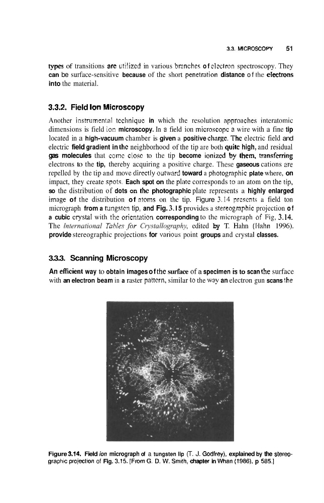
image of the distribution o f ntorns on the tip. Figure 3.14 prcscnts a field ton
micrograph from a hungstcn tip, and Fig. 3. I 5 provides a stereogmphic projection o f
a cubic crystal with the oricntation corresponding to the micrograph of Fig, 3.14.
The Irt~w~!ional TahIes jhr Cty~!d/opnphy, edited by T. Hahn (Ihhn 1996).
provide stereographic projections for various point groups and crystal classes.
3.3.3. Scanning Microscopy
An eficient way to obtain images o f thc surface of a specimen is to scan the surface
with an electron beam in a raster pattern, similar to the way an electron gun scans the
Figure 3.M. Field ion micrograph of a tungsten lip (T. J. Godfrey), explained by the stereographc projectton of Fig. 3.t5. IFrom G, D. W. Smith, chapler in Whan (?936), p 585.1
51
types of transitions arc utilized in various branches o f clcctron spectroscopy. They
can be surface-sensitive because of the short penetration distance o f the electrons
into the material.
3.3.2. Field Ion Microscopy
Another instrumental tcchnique in which the resolution approaches interatomic
dimensions is field ion microscopy. In a field ion microscopc o wire with a fine tip
located in a high-vacuum chamber is given a positive chargc. The electric field and
electric field gradient in thc neighborhood of the tip are both quitc high, and residual
gas molecules that comc close to the tip become ionized by them, transferaing
electrons to the tip, thereby acquiring a positive charge. Thew gaseous cations are
repelled by the tip and move directly outward toward a photogrnphic plate where, on
impact, they create spots. Each spot on the plate corresponds to an atom on the tip,
so the distribution of dots on the photographic plate represents a highly enlarged
image of the distribution o f ntorns on the tip. Figure 3.14 prcscnts a field ton
micrograph from a hungstcn tip, and Fig. 3. I 5 provides a stereogmphic projection o f
a cubic crystal with the oricntation corresponding to the micrograph of Fig, 3.14.
The Irt~w~!ional TahIes jhr Cty~!d/opnphy, edited by T. Hahn (Ihhn 1996).
provide stereographic projections for various point groups and crystal classes.
3.3.3. Scanning Microscopy
An eficient way to obtain images o f thc surface of a specimen is to scan the surface
with an electron beam in a raster pattern, similar to the way an electron gun scans the
Figure 3.M. Field ion micrograph of a tungsten lip (T. J. Godfrey), explained by the stereographc projectton of Fig. 3.t5. IFrom G, D. W. Smith, chapler in Whan (?936), p 585.1