

320
BIOLOGICAL MATERIALS
Pyrimidine Base
Purine Base
“2
I
0
II
t
t
t
t
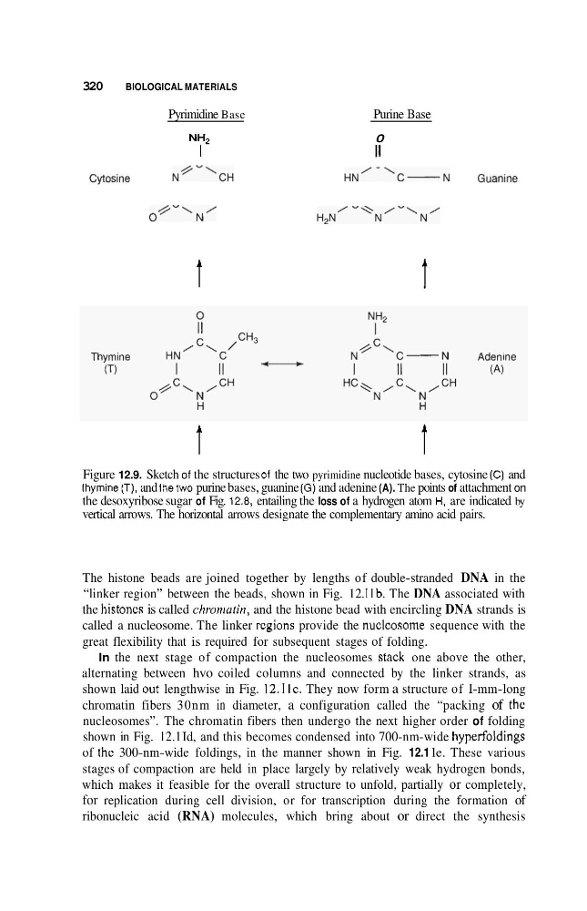
Figure 12.9. Sketch of the structures 01 the two pyrimidine nucleotide bases, cytosine (C) and
thymine (T). and thetwo purine bases, guanine ( G ) and adenine (A). The points of attachment on
the desoxyribose sugar of Fig. 12.8, entailing the loss of a hydrogen atom H. are indicated by
vertical arrows. The horizontal arrows designate the complementary amino acid pairs.
The histone beads are joined together by lengths of double-stranded DNA in the
“linker region” between the beads, shown in Fig. 12. I Ib. The DNA associated with
the histoncs is called chmniafin, and the histone bead with encircling DNA strands is
called a nucleosome. The linker rcgions provide the nuclcosome sequence with the
great flexibility that is required for subsequent stages of folding.
In the next stage of compaction the nucleosomes skick one above the other,
alternating between hvo coiled columns and connected by the linker strands, as
shown laid out lengthwise in Fig. 12. I IC. They now form a structure of I-mm-long
chromatin fibers 30nm in diameter, a configuration called the “packing of the
nucleosomes”. The chromatin fibers then undergo the next higher order of folding
shown in Fig. 12.1 Id, and this becomes condensed into 700-nm-wide hyperfoldings
of thc 300-nm-wide foldings, in the manner shown in Fig. 12.1 le. These various
stages of compaction are held in place largely by relatively weak hydrogen bonds,
which makes it feasible for the overall structure to unfold, partially or completely,
for replication during cell division, or for transcription during the formation of
ribonucleic acid (RNA) molecules, which bring about or direct the synthesis
BIOLOGICAL MATERIALS
Pyrimidine Base
Purine Base
“2
I
0
II
t
t
t
t
Figure 12.9. Sketch of the structures 01 the two pyrimidine nucleotide bases, cytosine (C) and
thymine (T). and thetwo purine bases, guanine ( G ) and adenine (A). The points of attachment on
the desoxyribose sugar of Fig. 12.8, entailing the loss of a hydrogen atom H. are indicated by
vertical arrows. The horizontal arrows designate the complementary amino acid pairs.
The histone beads are joined together by lengths of double-stranded DNA in the
“linker region” between the beads, shown in Fig. 12. I Ib. The DNA associated with
the histoncs is called chmniafin, and the histone bead with encircling DNA strands is
called a nucleosome. The linker rcgions provide the nuclcosome sequence with the
great flexibility that is required for subsequent stages of folding.
In the next stage of compaction the nucleosomes skick one above the other,
alternating between hvo coiled columns and connected by the linker strands, as
shown laid out lengthwise in Fig. 12. I IC. They now form a structure of I-mm-long
chromatin fibers 30nm in diameter, a configuration called the “packing of the
nucleosomes”. The chromatin fibers then undergo the next higher order of folding
shown in Fig. 12.1 Id, and this becomes condensed into 700-nm-wide hyperfoldings
of thc 300-nm-wide foldings, in the manner shown in Fig. 12.1 le. These various
stages of compaction are held in place largely by relatively weak hydrogen bonds,
which makes it feasible for the overall structure to unfold, partially or completely,
for replication during cell division, or for transcription during the formation of
ribonucleic acid (RNA) molecules, which bring about or direct the synthesis