

12.3. NUCLEIC ACIDS
319
Attachment
point for next
desoxyribose sugar
Nucleotide Base
H
0
I
0
H
Phosphate
Group
Attachment
point for next
H replaced by
phosphate group
OH lor RNA
Desoxyribose sugai
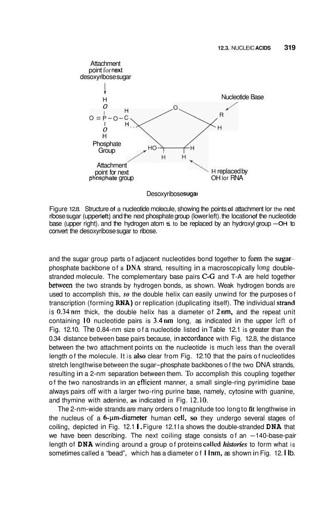
Figure 12.8. Structure of a nucleotide molecule, showing the points of attachment lor the next
ribose sugar (upper lelt) and the next phosphate group (lower left). the location of the nucleotide
base (upper right). and the hydrogen atom ti to be replaced by an hydroxyl group -OH to
convert the desoxyribose sugar to ribose.
and the sugar group parts o f adjacent nucleotides bond together to form the sugarphosphate backbone o f a DNA strand, resulting in a macroscopically 10116 doublestranded molecule. The complementary base pairs C-G and T-A are held together
between the two strands by hydrogen bonds, as shown. Weak hydrogen bonds are
used to accomplish this, so the double helix can easily unwind for the purposes o f
transcription (forming RNA) or replication (duplicating itself). The individual strand
is 0.34nm thick, the double helix has a diameter o f 2nm. and the repeat unit
containing I O nucleotide pairs is 3.4nm long, as indicated in the upper let? o f
Fig. 12.10. The 0.84-nm size o f a nucleotide listed in Table 12.1 is greater than the
0.34 distance between base pairs because, in accordance with Fig. 12.8, the distance
between the two attachment points on the nucleotide is much less than the overall
length o f the molecule. It i s also clear from Fig. 12.10 that the pairs o f nucleotides
stretch lengthwise between the sugar-phosphate backbones o f the two DNA strands,
resulting in a 2-nm separation between them. To accomplish this coupling together
o f the two nanostrands in an eflicient manner, a small single-ring pyrimidine base
always pairs off with a larger two-ring purine base, namely, cytosine with guanine,
and thymine with adenine, as indicated in Fig. 12.10.
The 2-nm-wide strands are many orders o f magnitude too long to fit lengthwise in
the nucleus of a 6-pm-diameter human cell, so they undergo several stages of
coiling, depicted in Fig. 12.1 I. Figure 12.1 l a shows the double-stranded DNA that
we have been describing. The next coiling stage consists o f an -140-base-pair
length of DNA winding around a group o f proteins called histories to form what i s
sometimes called a “bead”, which has a diameter o f I I nm, as shown in Fig. 12. I I b.
319
Attachment
point for next
desoxyribose sugar
Nucleotide Base
H
0
I
0
H
Phosphate
Group
Attachment
point for next
H replaced by
phosphate group
OH lor RNA
Desoxyribose sugai
Figure 12.8. Structure of a nucleotide molecule, showing the points of attachment lor the next
ribose sugar (upper lelt) and the next phosphate group (lower left). the location of the nucleotide
base (upper right). and the hydrogen atom ti to be replaced by an hydroxyl group -OH to
convert the desoxyribose sugar to ribose.
and the sugar group parts o f adjacent nucleotides bond together to form the sugarphosphate backbone o f a DNA strand, resulting in a macroscopically 10116 doublestranded molecule. The complementary base pairs C-G and T-A are held together
between the two strands by hydrogen bonds, as shown. Weak hydrogen bonds are
used to accomplish this, so the double helix can easily unwind for the purposes o f
transcription (forming RNA) or replication (duplicating itself). The individual strand
is 0.34nm thick, the double helix has a diameter o f 2nm. and the repeat unit
containing I O nucleotide pairs is 3.4nm long, as indicated in the upper let? o f
Fig. 12.10. The 0.84-nm size o f a nucleotide listed in Table 12.1 is greater than the
0.34 distance between base pairs because, in accordance with Fig. 12.8, the distance
between the two attachment points on the nucleotide is much less than the overall
length o f the molecule. It i s also clear from Fig. 12.10 that the pairs o f nucleotides
stretch lengthwise between the sugar-phosphate backbones o f the two DNA strands,
resulting in a 2-nm separation between them. To accomplish this coupling together
o f the two nanostrands in an eflicient manner, a small single-ring pyrimidine base
always pairs off with a larger two-ring purine base, namely, cytosine with guanine,
and thymine with adenine, as indicated in Fig. 12.10.
The 2-nm-wide strands are many orders o f magnitude too long to fit lengthwise in
the nucleus of a 6-pm-diameter human cell, so they undergo several stages of
coiling, depicted in Fig. 12.1 I. Figure 12.1 l a shows the double-stranded DNA that
we have been describing. The next coiling stage consists o f an -140-base-pair
length of DNA winding around a group o f proteins called histories to form what i s
sometimes called a “bead”, which has a diameter o f I I nm, as shown in Fig. 12. I I b.