

31 2
BIOLOGICAL MATERIALS
Another common way to detcrrnine the size o f a biological molecule is to obscrve
i t using an electron rnicmscopc, which can provide images called e'cctma micrngmphs taken Frclni various molecular orientations. This approach IS especially uscfuI
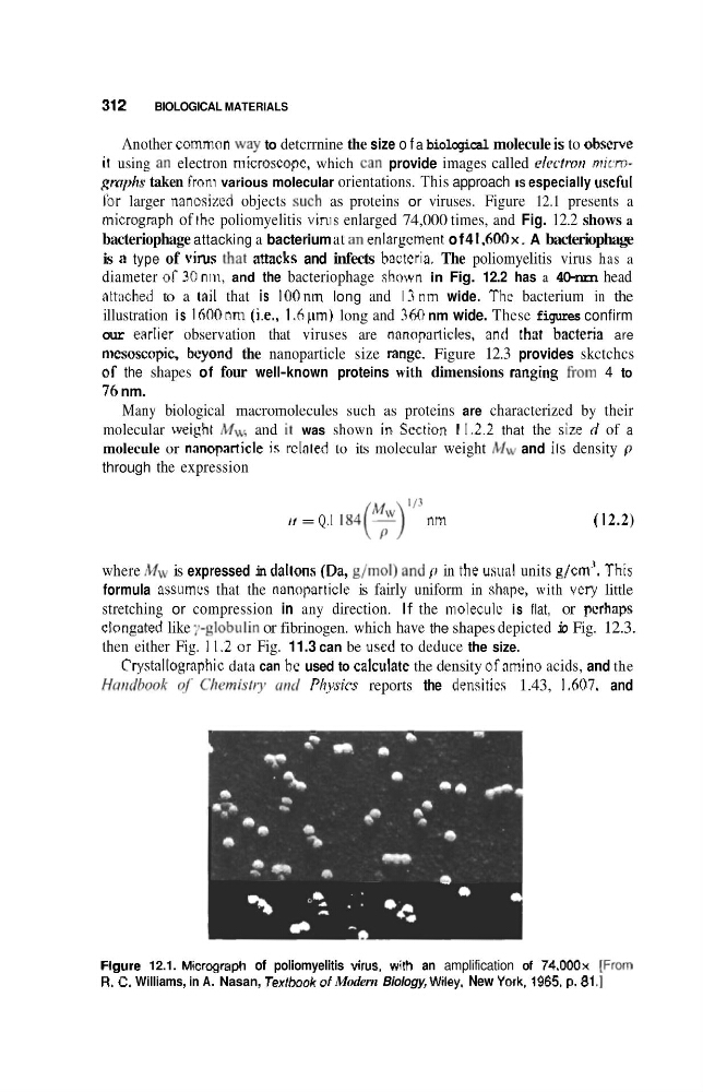
Ibr larger nanosizvd objects such as proteins or viruses. Figure 12.1 presents a
micrograph of rhc poliomyelitis vlnis enlarged 74,000 times, and Fig. 12.2 shows a
bacteriophage attacking a bacterium at an enlargcrnent of4 1 . 6 0 0 ~ .
A bacteriophage
is B type of virus !ha! amcks and infects bacrcria. The poliomyelitis virus has a
diameter 06- 30nii1, and the bacteriophage shown in Fig. 12.2 has a 40-nrn head
attached to a tail that is 100nm long and 13nm wide. Thc bacterium in the
illustration is 1600 nm (i.e., 1 .h pm) long and 360 nm wide. These figures confirm
our earlier observation that viruses are nnnoparticles, ant! that bacteria are
mcsoscopic. beyond the nanoparticle size rangc. Figure 12.3 provides skctchcs
06 the shapes of four well-known proteins with dimensions ranging From 4 to
7'h nm.
Many biological macromolecules such as proteins are characterized by their
molecular weight MI\, and in was shown in ScctEon I I .2.2 that the size d of a
molecule or nanopamicle is d a t e d to its molecular weight MW and ils density p
through the expression
if = Q.l 1 8 4 ( $ ) ' "
nm
( 1 2.2)
where M ~ v
is expressed in daItons (Qa, g/mol) and p in the usitnl units gJcm'. This
formula assums that the nanoparticle is fairly uniform in shape, with very little
stretching or compression in any direction. If the moleculv is flat, or pcrhaps
elongated like ;.-gIo,buIin or fibrinogen. which have the shapes depicted io Fig. 12.3.
then either Fig. 11.2 or Fig. 11.3 can be used to deduce the size.
Cryshltogmphic data can he used to calculate the density ofamino acids, and the
HmdlmA' of i'htvnisfty urd PJzysies reports the densitics 1.43, 1.607. and
Figure 12.1. Micrwraph of poliomyelitis virus, wilh an amplification of 74,000~ [FrQom
R. C. Williams, in A. Nasan, Textbook d Modern Biology, Wiley, New York, 1965, p. 81 .I
BIOLOGICAL MATERIALS
Another common way to detcrrnine the size o f a biological molecule is to obscrve
i t using an electron rnicmscopc, which can provide images called e'cctma micrngmphs taken Frclni various molecular orientations. This approach IS especially uscfuI
Ibr larger nanosizvd objects such as proteins or viruses. Figure 12.1 presents a
micrograph of rhc poliomyelitis vlnis enlarged 74,000 times, and Fig. 12.2 shows a
bacteriophage attacking a bacterium at an enlargcrnent of4 1 . 6 0 0 ~ .
A bacteriophage
is B type of virus !ha! amcks and infects bacrcria. The poliomyelitis virus has a
diameter 06- 30nii1, and the bacteriophage shown in Fig. 12.2 has a 40-nrn head
attached to a tail that is 100nm long and 13nm wide. Thc bacterium in the
illustration is 1600 nm (i.e., 1 .h pm) long and 360 nm wide. These figures confirm
our earlier observation that viruses are nnnoparticles, ant! that bacteria are
mcsoscopic. beyond the nanoparticle size rangc. Figure 12.3 provides skctchcs
06 the shapes of four well-known proteins with dimensions ranging From 4 to
7'h nm.
Many biological macromolecules such as proteins are characterized by their
molecular weight MI\, and in was shown in ScctEon I I .2.2 that the size d of a
molecule or nanopamicle is d a t e d to its molecular weight MW and ils density p
through the expression
if = Q.l 1 8 4 ( $ ) ' "
nm
( 1 2.2)
where M ~ v
is expressed in daItons (Qa, g/mol) and p in the usitnl units gJcm'. This
formula assums that the nanoparticle is fairly uniform in shape, with very little
stretching or compression in any direction. If the moleculv is flat, or pcrhaps
elongated like ;.-gIo,buIin or fibrinogen. which have the shapes depicted io Fig. 12.3.
then either Fig. 11.2 or Fig. 11.3 can be used to deduce the size.
Cryshltogmphic data can he used to calculate the density ofamino acids, and the
HmdlmA' of i'htvnisfty urd PJzysies reports the densitics 1.43, 1.607. and
Figure 12.1. Micrwraph of poliomyelitis virus, wilh an amplification of 74,000~ [FrQom
R. C. Williams, in A. Nasan, Textbook d Modern Biology, Wiley, New York, 1965, p. 81 .I