

12.2. BIOLOGICAL BUILDING BLOCKS
311
molecule is a double nanowire, hvo nucleotide nanowires twisted around each with a
repeat unit every 3.4nm, and a diameter of 2nm. This long double-stranded
nanowire also undergocs systcmatic twistings and turnings to become con~pacted
into a chromosome about 6 pm long and I .4 pm wide. The chromoson~e itself i s not
small enough to he a nanoparticlc; rather, it is in the mesoscopic range of size.
To gain some additional perspective about the overall scope of nanometer-range
sizes involved in the buildup of hiological stnictures. let us consider the human
tendon as a typical structure (Tirrcll 1994). The function of a tendon i s to attach a
muscle to a bone. From the viewpoint of biology, the fundamental building block of
a tendon is the assemblage of amino acids (0.6 nm) that form the gelatinlike protein
called collagen (I nm), which coils into a triplc hclix (2nm). There follows a
threefold sequence of fiberlike or fibrillar nanostructures: a microfibril (3.5 nm), a
subfibril (10-20nm). and a fibril itself (50-500nm). The final two steps in the
buildup, specifically, the cluster o f fibers called a /irscic/t. (50-300pm) and the
tendon itself (10-50 cm). are far beyond thc nanometcr range of sizes. The fascicle is
considered mesoscopic and the tendon, macroscopic in size. Since the smallest
amino acid glycine, is -0.42 nm in size. and some vinises reach 200nm. it seems
appropriate to define a biological nanosmlchire as k i n g in the nominal range from
0.5 to 200nm. With this in mind, the present chapter focuscs on nanonieter-size
constituents of biological materials. In addition, we also comment on some special
cases in which artificially constructed nanostnicturcs are o f importance in biology.
See Gross ( I 999) for some additional discussions of biological nanoshuctures.
12.2. BIOLOGICAL BUILDING BLOCKS
12.2.1. Sizes of Building Blocks and Nanostructures
There are a number of ways to determine or estimate the size panmcters d of the
fundamental biological building blocks, which are amino acids for protcins and
nucleotides for DNA. I f the crystal structure is known for the building-block
molecule, and there are n molecules in the crystallographic unit cell. thcn one can
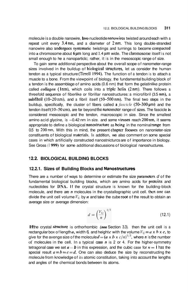
divide the unit cell volume Vu by II and take the cube r m t o f the result to obtain an
average size or average dimension:
(12.1)
If the crystal structure is orthorhombic (see Section 3.3). then the unit cell is a
rectangular box o f length a, width 6, and height c with the volume Vu = u x h x c, to
give for the avenge size o f the molecule d=(a x h x c/n)"', where n is thc number
of molecules in the cell. In a typical case n is 2 or 4. For the higher-symmetry
tetragonal case we set a = b in this expression, and the cubic case for n = I has the
special result a = h = c = d . One can also deduce the size by reconstructing the
molecule from knowledge of its atomic constitution, taking into account the lengths
and angles of the chemical bonds between its atoms.
311
molecule is a double nanowire, hvo nucleotide nanowires twisted around each with a
repeat unit every 3.4nm, and a diameter of 2nm. This long double-stranded
nanowire also undergocs systcmatic twistings and turnings to become con~pacted
into a chromosome about 6 pm long and I .4 pm wide. The chromoson~e itself i s not
small enough to he a nanoparticlc; rather, it is in the mesoscopic range of size.
To gain some additional perspective about the overall scope of nanometer-range
sizes involved in the buildup of hiological stnictures. let us consider the human
tendon as a typical structure (Tirrcll 1994). The function of a tendon i s to attach a
muscle to a bone. From the viewpoint of biology, the fundamental building block of
a tendon is the assemblage of amino acids (0.6 nm) that form the gelatinlike protein
called collagen (I nm), which coils into a triplc hclix (2nm). There follows a
threefold sequence of fiberlike or fibrillar nanostructures: a microfibril (3.5 nm), a
subfibril (10-20nm). and a fibril itself (50-500nm). The final two steps in the
buildup, specifically, the cluster o f fibers called a /irscic/t. (50-300pm) and the
tendon itself (10-50 cm). are far beyond thc nanometcr range of sizes. The fascicle is
considered mesoscopic and the tendon, macroscopic in size. Since the smallest
amino acid glycine, is -0.42 nm in size. and some vinises reach 200nm. it seems
appropriate to define a biological nanosmlchire as k i n g in the nominal range from
0.5 to 200nm. With this in mind, the present chapter focuscs on nanonieter-size
constituents of biological materials. In addition, we also comment on some special
cases in which artificially constructed nanostnicturcs are o f importance in biology.
See Gross ( I 999) for some additional discussions of biological nanoshuctures.
12.2. BIOLOGICAL BUILDING BLOCKS
12.2.1. Sizes of Building Blocks and Nanostructures
There are a number of ways to determine or estimate the size panmcters d of the
fundamental biological building blocks, which are amino acids for protcins and
nucleotides for DNA. I f the crystal structure is known for the building-block
molecule, and there are n molecules in the crystallographic unit cell. thcn one can
divide the unit cell volume Vu by II and take the cube r m t o f the result to obtain an
average size or average dimension:
(12.1)
If the crystal structure is orthorhombic (see Section 3.3). then the unit cell is a
rectangular box o f length a, width 6, and height c with the volume Vu = u x h x c, to
give for the avenge size o f the molecule d=(a x h x c/n)"', where n is thc number
of molecules in the cell. In a typical case n is 2 or 4. For the higher-symmetry
tetragonal case we set a = b in this expression, and the cubic case for n = I has the
special result a = h = c = d . One can also deduce the size by reconstructing the
molecule from knowledge of its atomic constitution, taking into account the lengths
and angles of the chemical bonds between its atoms.