

11.5. SUPRAMOLECULAR STRUCTURES
307
lnleraction with
Ligand rnokcules:
sugar and allgopeptide
PEG laver inhibits non-soecrfic
adsorpiion of protelns '
BiodegradaWe surface
4
Functionnliration of
polytacwh surface
Functional group:
Acetal
Hydrophilic segment
double bond
Dialysis
in water
- t
r
. L .
Surface
I, - -
j
+ ,
c:
Micelle foormatlon modification Cork-po l yrnerized micelle
with aldehyde groups
on Ihe surface
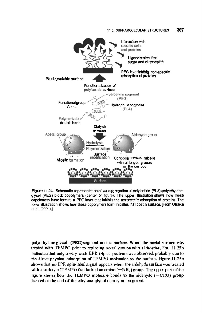
Figure 11.24. Schematic representation of an aggregation of polylactide (PlA)/polyelhyleneglycol (PEG) block copolymers (center of fgure). The upper illustration shows how Ihese
copolymers have tormed a PEG layer that inhibits the nonspecific adsorption of proteins. The
lower illustration shows how these copolymers form micelles thal coat a surface. [From Otsuka
et al. (2001).]
polyethylene glycol (PEG) segment on the surface. When the acetal surface was
?mated with TEMPO prior to replacing acetal groups with aldehydes, Fig. 11.2%
indicates that only a v c p weak EPR triplet spectrum was obcrved, probably due to
the direct physical adsorption of TEMPO molecules on tlic surface. Figure 1 1 . 2 5 ~
shows that RO EPR spin-labcl signal appears when the aldchydc surtice was treated
with a variety o f TEMPO lhnt lacked an amino ( -NH2) gmup. The upper part o f the
figure shows how the TEMPO molecule bonds to the aldehydc (-CHO) group
located at the end of tlic ctliylene glycol copolymer segment.
307
lnleraction with
Ligand rnokcules:
sugar and allgopeptide
PEG laver inhibits non-soecrfic
adsorpiion of protelns '
BiodegradaWe surface
4
Functionnliration of
polytacwh surface
Functional group:
Acetal
Hydrophilic segment
double bond
Dialysis
in water
- t
r
. L .
Surface
I, - -
j
+ ,
c:
Micelle foormatlon modification Cork-po l yrnerized micelle
with aldehyde groups
on Ihe surface
Figure 11.24. Schematic representation of an aggregation of polylactide (PlA)/polyelhyleneglycol (PEG) block copolymers (center of fgure). The upper illustration shows how Ihese
copolymers have tormed a PEG layer that inhibits the nonspecific adsorption of proteins. The
lower illustration shows how these copolymers form micelles thal coat a surface. [From Otsuka
et al. (2001).]
polyethylene glycol (PEG) segment on the surface. When the acetal surface was
?mated with TEMPO prior to replacing acetal groups with aldehydes, Fig. 11.2%
indicates that only a v c p weak EPR triplet spectrum was obcrved, probably due to
the direct physical adsorption of TEMPO molecules on tlic surface. Figure 1 1 . 2 5 ~
shows that RO EPR spin-labcl signal appears when the aldchydc surtice was treated
with a variety o f TEMPO lhnt lacked an amino ( -NH2) gmup. The upper part o f the
figure shows how the TEMPO molecule bonds to the aldehydc (-CHO) group
located at the end of tlic ctliylene glycol copolymer segment.