

66
3 Fundamentals of DET-Type Bioelectrocatalysis
Fig. 3.2 Schematic of
DET-type bioelectrocatalysis
of FAD-GOD and
FAD-GDH wired with
platinum or gold
nanoclusters at the electrode
surface
glucose
interfacial electron transfer
electrode
gluconate
FAD
Pt/Au nanoclusters
[56, 57]. Pt nanoclusters were grown enzymatic reaction of the enzyme with PtCl 6
2−
as an electron acceptor and were enable electrical contact between the enzyme and
an electrode. Single-walled carbon nanotubes are suggested to accelerate DET-type
bioelectrocatalysis of FAD-dependent GDH [58]. Enzymatically implanted platinum
nanoclusters on porous gold electrodes also worked well as scaffolds for DET-type
bioelectrocatalysis of FAD-dependent GDH [59].
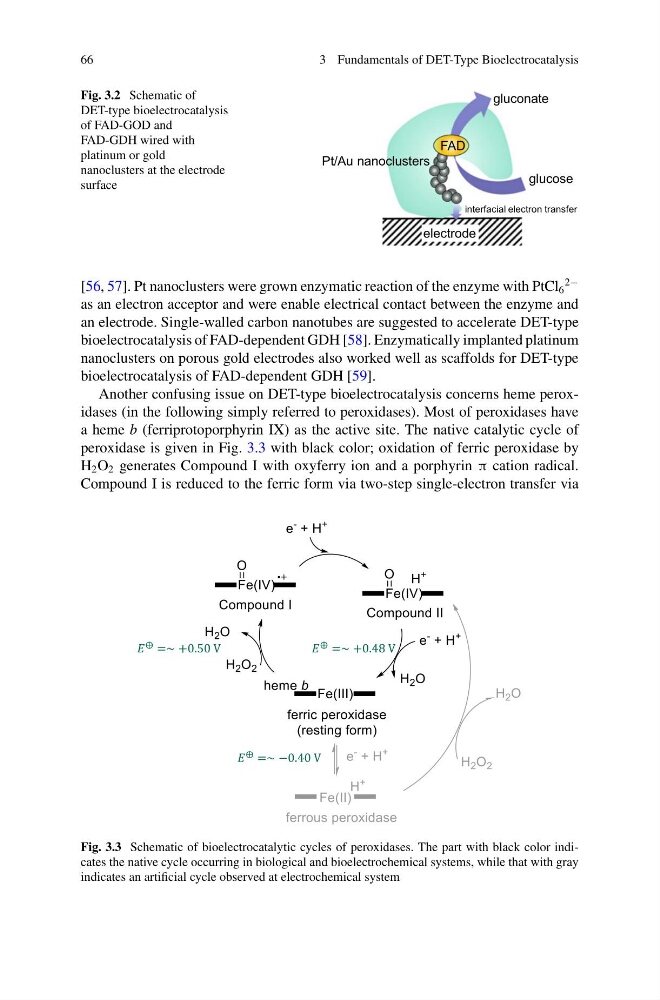
Another confusing issue on DET-type bioelectrocatalysis concerns heme peroxidases (in the following simply referred to peroxidases). Most of peroxidases have
a heme b (ferriprotoporphyrin IX) as the active site. The native catalytic cycle of
peroxidase is given in Fig. 3.3 with black color; oxidation of ferric peroxidase by
H 2 O 2 generates Compound I with oxyferry ion and a porphyrin π cation radical.
Compound I is reduced to the ferric form via two-step single-electron transfer via
Fig. 3.3 Schematic of bioelectrocatalytic cycles of peroxidases. The part with black color indicates the native cycle occurring in biological and bioelectrochemical systems, while that with gray
indicates an artificial cycle observed at electrochemical system
3 Fundamentals of DET-Type Bioelectrocatalysis
Fig. 3.2 Schematic of
DET-type bioelectrocatalysis
of FAD-GOD and
FAD-GDH wired with
platinum or gold
nanoclusters at the electrode
surface
glucose
interfacial electron transfer
electrode
gluconate
FAD
Pt/Au nanoclusters
[56, 57]. Pt nanoclusters were grown enzymatic reaction of the enzyme with PtCl 6
2−
as an electron acceptor and were enable electrical contact between the enzyme and
an electrode. Single-walled carbon nanotubes are suggested to accelerate DET-type
bioelectrocatalysis of FAD-dependent GDH [58]. Enzymatically implanted platinum
nanoclusters on porous gold electrodes also worked well as scaffolds for DET-type
bioelectrocatalysis of FAD-dependent GDH [59].
Another confusing issue on DET-type bioelectrocatalysis concerns heme peroxidases (in the following simply referred to peroxidases). Most of peroxidases have
a heme b (ferriprotoporphyrin IX) as the active site. The native catalytic cycle of
peroxidase is given in Fig. 3.3 with black color; oxidation of ferric peroxidase by
H 2 O 2 generates Compound I with oxyferry ion and a porphyrin π cation radical.
Compound I is reduced to the ferric form via two-step single-electron transfer via
Fig. 3.3 Schematic of bioelectrocatalytic cycles of peroxidases. The part with black color indicates the native cycle occurring in biological and bioelectrochemical systems, while that with gray
indicates an artificial cycle observed at electrochemical system