

5.2 Enzyme Trimming
95
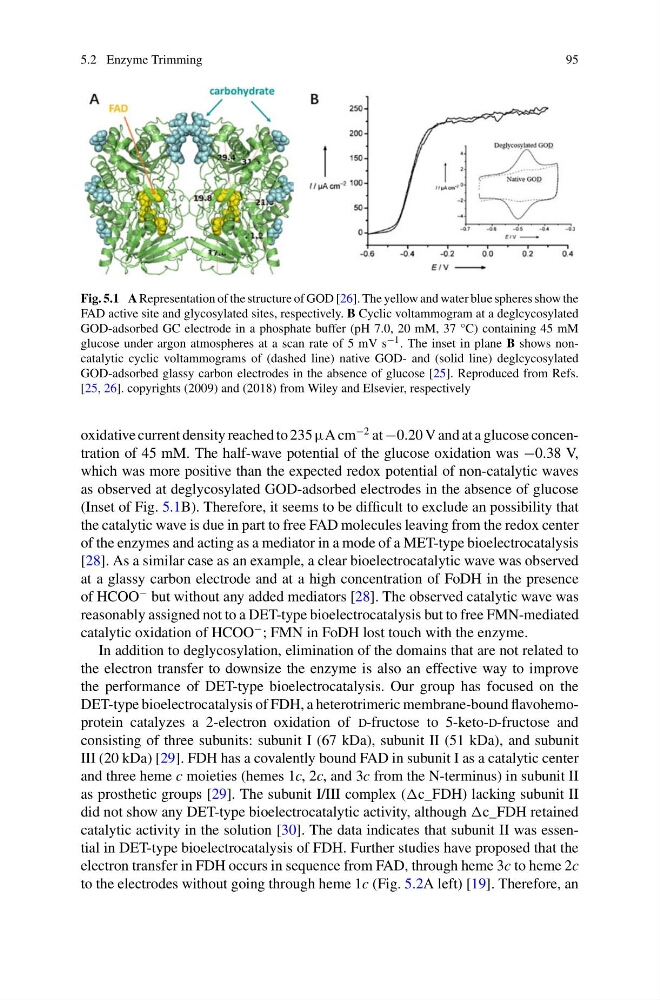
Fig. 5.1 A Representation of the structure of GOD [26]. The yellow and water blue spheres show the
FAD active site and glycosylated sites, respectively. B Cyclic voltammogram at a deglcycosylated
GOD-adsorbed GC electrode in a phosphate buffer (pH 7.0, 20 mM, 37 °C) containing 45 mM
glucose under argon atmospheres at a scan rate of 5 mV s −1 . The inset in plane B shows noncatalytic cyclic voltammograms of (dashed line) native GOD- and (solid line) deglcycosylated
GOD-adsorbed glassy carbon electrodes in the absence of glucose [25]. Reproduced from Refs.
[25, 26]. copyrights (2009) and (2018) from Wiley and Elsevier, respectively
oxidative current density reached to 235 µA cm
−2 at −0.20 V and at a glucose concentration of 45 mM. The half-wave potential of the glucose oxidation was −0.38 V,
which was more positive than the expected redox potential of non-catalytic waves
as observed at deglycosylated GOD-adsorbed electrodes in the absence of glucose
(Inset of Fig. 5.1B). Therefore, it seems to be difficult to exclude an possibility that
the catalytic wave is due in part to free FAD molecules leaving from the redox center
of the enzymes and acting as a mediator in a mode of a MET-type bioelectrocatalysis
[28]. As a similar case as an example, a clear bioelectrocatalytic wave was observed
at a glassy carbon electrode and at a high concentration of FoDH in the presence
of HCOO
− but without any added mediators [28]. The observed catalytic wave was
reasonably assigned not to a DET-type bioelectrocatalysis but to free FMN-mediated
catalytic oxidation of HCOO
− ; FMN in FoDH lost touch with the enzyme.
In addition to deglycosylation, elimination of the domains that are not related to
the electron transfer to downsize the enzyme is also an effective way to improve
the performance of DET-type bioelectrocatalysis. Our group has focused on the
DET-type bioelectrocatalysis of FDH, a heterotrimeric membrane-bound flavohemoprotein catalyzes a 2-electron oxidation of d-fructose to 5-keto-d-fructose and
consisting of three subunits: subunit I (67 kDa), subunit II (51 kDa), and subunit
III (20 kDa) [29]. FDH has a covalently bound FAD in subunit I as a catalytic center
and three heme c moieties (hemes 1c, 2c, and 3c from the N-terminus) in subunit II
as prosthetic groups [29]. The subunit I/III complex (c_FDH) lacking subunit II
did not show any DET-type bioelectrocatalytic activity, although c_FDH retained
catalytic activity in the solution [30]. The data indicates that subunit II was essential in DET-type bioelectrocatalysis of FDH. Further studies have proposed that the
electron transfer in FDH occurs in sequence from FAD, through heme 3c to heme 2c
to the electrodes without going through heme 1c (Fig. 5.2A left) [19]. Therefore, an
95
Fig. 5.1 A Representation of the structure of GOD [26]. The yellow and water blue spheres show the
FAD active site and glycosylated sites, respectively. B Cyclic voltammogram at a deglcycosylated
GOD-adsorbed GC electrode in a phosphate buffer (pH 7.0, 20 mM, 37 °C) containing 45 mM
glucose under argon atmospheres at a scan rate of 5 mV s −1 . The inset in plane B shows noncatalytic cyclic voltammograms of (dashed line) native GOD- and (solid line) deglcycosylated
GOD-adsorbed glassy carbon electrodes in the absence of glucose [25]. Reproduced from Refs.
[25, 26]. copyrights (2009) and (2018) from Wiley and Elsevier, respectively
oxidative current density reached to 235 µA cm
−2 at −0.20 V and at a glucose concentration of 45 mM. The half-wave potential of the glucose oxidation was −0.38 V,
which was more positive than the expected redox potential of non-catalytic waves
as observed at deglycosylated GOD-adsorbed electrodes in the absence of glucose
(Inset of Fig. 5.1B). Therefore, it seems to be difficult to exclude an possibility that
the catalytic wave is due in part to free FAD molecules leaving from the redox center
of the enzymes and acting as a mediator in a mode of a MET-type bioelectrocatalysis
[28]. As a similar case as an example, a clear bioelectrocatalytic wave was observed
at a glassy carbon electrode and at a high concentration of FoDH in the presence
of HCOO
− but without any added mediators [28]. The observed catalytic wave was
reasonably assigned not to a DET-type bioelectrocatalysis but to free FMN-mediated
catalytic oxidation of HCOO
− ; FMN in FoDH lost touch with the enzyme.
In addition to deglycosylation, elimination of the domains that are not related to
the electron transfer to downsize the enzyme is also an effective way to improve
the performance of DET-type bioelectrocatalysis. Our group has focused on the
DET-type bioelectrocatalysis of FDH, a heterotrimeric membrane-bound flavohemoprotein catalyzes a 2-electron oxidation of d-fructose to 5-keto-d-fructose and
consisting of three subunits: subunit I (67 kDa), subunit II (51 kDa), and subunit
III (20 kDa) [29]. FDH has a covalently bound FAD in subunit I as a catalytic center
and three heme c moieties (hemes 1c, 2c, and 3c from the N-terminus) in subunit II
as prosthetic groups [29]. The subunit I/III complex (c_FDH) lacking subunit II
did not show any DET-type bioelectrocatalytic activity, although c_FDH retained
catalytic activity in the solution [30]. The data indicates that subunit II was essential in DET-type bioelectrocatalysis of FDH. Further studies have proposed that the
electron transfer in FDH occurs in sequence from FAD, through heme 3c to heme 2c
to the electrodes without going through heme 1c (Fig. 5.2A left) [19]. Therefore, an