

(c) Incubate the slides in 80% and 90% ethanol, 2 min each.
(d) Incubate the slides two times in xylene, 5 min each.
(e) Mount the specimens in mounting medium.
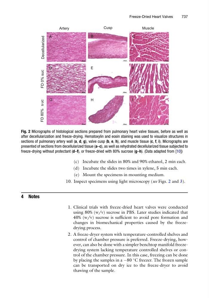
10. Inspect specimens using light microscopy (see Figs. 2 and 3).
4 Notes
1. Clinical trials with freeze-dried heart valves were conducted
using 80% (w/v) sucrose in PBS. Later studies indicated that
40% (w/v) sucrose is sufficient to avoid pore formation and
changes in biomechanical properties caused by the freezedrying process.
2. A freeze-dryer system with temperature-controlled shelves and
control of chamber pressure is preferred. Freeze-drying, however, can also be done with a simpler benchtop manifold freezedrying system lacking temperature controlled shelves or control of the chamber pressure. In this case, freezing can be done
by placing the samples in a À80
C freezer. The frozen sample
can be transported on dry ice to the freeze-dryer to avoid
thawing of the sample.
Decellularized
FD 0% suc
FD 80% s uc
Artery
Cusp
Muscle
A
B
C
D
E
F
G
H
I
Fig. 2 Micrographs of histological sections prepared from pulmonary heart valve tissues, before as well as
after decellularization and freeze-drying. Hematoxylin and eosin staining was used to visualize structures in
sections of pulmonary artery wall (a, d, g), valve cusp (b, e, h), and muscle tissue (c, f, i). Micrographs are
presented of sections from decellularized tissue (a–c), as well as rehydrated decellularized tissue subjected to
freeze-drying without protectant (d–f), or freeze-dried with 80% sucrose (g–h). (Data adapted from [10])
Freeze-Dried Heart Valves
737