

5. Cryo-dewar large/small (e.g., Archive Dewar, Biosystem
36 and Biorack 6000, Statebourne.com).
6. Cold-protective gloves (cryo-gloves) and protective glasses.
7. Liquid nitrogen.
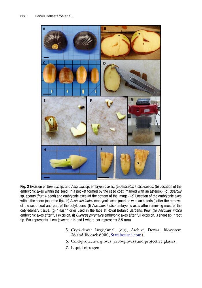
Fig. 2 Excision of Quercus sp. and Aesculus sp. embryonic axes. (a) Aesculus indica seeds. (b) Location of the
embryonic axes within the seed, in a pocket formed by the seed coat (marked with an asterisk). (c) Quercus
sp. acorns (fruit + seed) and embryonic axes (at the bottom of the image). (d) Location of the embryonic axes
within the acorn (near the tip). (e) Aesculus indica embryonic axes (marked with an asterisk) after the removal
of the seed coat and part of the cotyledons. (f) Aesculus indica embryonic axes after removing most of the
cotyledonary tissue. (g) “Flash” drier used in the labs at Royal Botanic Gardens, Kew. (h) Aesculus indica
embryonic axes after full excision. (i) Quercus pyrenaica embryonic axes after full excision. s shoot tip, r root
tip. Bar represents 1 cm (except in h and i where bar represents 2.5 mm)
668
Daniel Ballesteros et al.