

umbilical vein endothelial cells were added to each tissue sample under physiological cell culture conditions in non-tissue
culture tubes (to minimize cell migration from the tissues).
There were no statistically significant viability differences comparing endothelial cell seeded decellularized vessels and ice-free
preserved tissue engineered blood vessels that were not decellularized after two, four, or eight washes in cell culture
[12]. The endothelial cells formed confluent monolayers on
all surfaces of the washed tissues, regardless of the number of
post-rewarming washes [12], demonstrating that any residual
cryoprotectants present were not cytotoxic. In parallel xenograft tissue studies, we have also demonstrated that viability of
human peripheral blood mononuclear cells is not impacted by
culture with rewarmed tissues washed 5Â with EC [23].
9. Addition of sugars to VS55 has a stabilizing effect during slow
cooling and warming of vitrified tissue samples. Comparison of
VS55 solutions supplemented with 0.1–0.6 M trehalose or
sucrose has demonstrated concentration-dependent inhibition
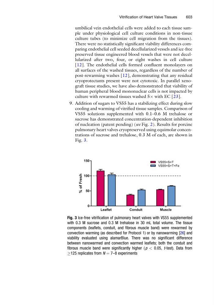
of nucleation (patent pending) (see Fig. 2). Results for porcine
pulmonary heart valves cryopreserved using equimolar concentrations of sucrose and trehalose, 0.3 M of each, are shown in
Fig. 3.
Fig. 3 Ice-free vitrification of pulmonary heart valves with VS55 supplemented
with 0.3 M sucrose and 0.3 M trehalose in 30 mL total volume. The tissue
components (leaflets, conduit, and fibrous muscle band) were rewarmed by
convection warming (as described for Protocol 1) or by nanowarming [26] and
viability evaluated using alamarBlue. There was no significant difference
between nanowarmed and convection warmed leaflets; both the conduit and
fibrous muscle band were significantly higher ( p < 0.05, t-test). Data from
!125 replicates from N ¼ 7–8 experiments
Vitrification of Heart Valve Tissues
603
culture tubes (to minimize cell migration from the tissues).
There were no statistically significant viability differences comparing endothelial cell seeded decellularized vessels and ice-free
preserved tissue engineered blood vessels that were not decellularized after two, four, or eight washes in cell culture
[12]. The endothelial cells formed confluent monolayers on
all surfaces of the washed tissues, regardless of the number of
post-rewarming washes [12], demonstrating that any residual
cryoprotectants present were not cytotoxic. In parallel xenograft tissue studies, we have also demonstrated that viability of
human peripheral blood mononuclear cells is not impacted by
culture with rewarmed tissues washed 5Â with EC [23].
9. Addition of sugars to VS55 has a stabilizing effect during slow
cooling and warming of vitrified tissue samples. Comparison of
VS55 solutions supplemented with 0.1–0.6 M trehalose or
sucrose has demonstrated concentration-dependent inhibition
of nucleation (patent pending) (see Fig. 2). Results for porcine
pulmonary heart valves cryopreserved using equimolar concentrations of sucrose and trehalose, 0.3 M of each, are shown in
Fig. 3.
Fig. 3 Ice-free vitrification of pulmonary heart valves with VS55 supplemented
with 0.3 M sucrose and 0.3 M trehalose in 30 mL total volume. The tissue
components (leaflets, conduit, and fibrous muscle band) were rewarmed by
convection warming (as described for Protocol 1) or by nanowarming [26] and
viability evaluated using alamarBlue. There was no significant difference
between nanowarmed and convection warmed leaflets; both the conduit and
fibrous muscle band were significantly higher ( p < 0.05, t-test). Data from
!125 replicates from N ¼ 7–8 experiments
Vitrification of Heart Valve Tissues
603