

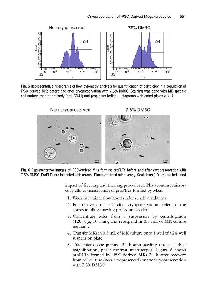
impact of freezing and thawing procedures. Phas-contrast microscopy allows visualization of proPLTs formed by MKs.
1. Work in laminar flow hood under sterile conditions.
2. For recovery of cells after cryopreservation, refer to the
corresponding thawing procedure section.
3. Concentrate MKs from a suspension by centrifugation
(120 Â g, 10 min), and resuspend in 0.5 mL of MK culture
medium.
4. Transfer MKs in 0.5 mL of MK culture onto 1 well of a 24-well
suspension plate.
5. Take microscope pictures 24 h after seeding the cells (40Â
magnification, phase-contrast microscope). Figure 6 shows
proPLTs formed by iPSC-derived MKs 24 h after recovery
from cell culture (non-cryopreserved) or after cryopreservation
with 7.5% DMSO.
Fig. 6 Representative images of iPSC-derived MKs forming proPLTs before and after cryopreservation with
7.5% DMSO. ProPLTs are indicated with arrows. Phase-contrast microscopy. Scale bars (10 μm) are indicated
Non-cryopreserved
n≥4
n ≥4
Count
7.5% DMSO
0
–33
10 2
10 3
PI-A
10 4
10 5
0 100 200 300 400 500 600 700
Count
–33
10 2
0 10 1
10 3
PI-A
10 4
10 5
0 100 200 300 400 500 600
Fig. 5 Representative histograms of flow cytometry analysis for quantification of polyploidy in a population of
iPSC-derived MKs before and after cryopreservation with 7.5% DMSO. Staining was done with MK-specific
cell surface marker antibody (anti-CD41) and propidium iodide. Histograms with gated ploidy n ! 4
Cryopreservation of iPSC-Derived Megakaryocytes
551
1. Work in laminar flow hood under sterile conditions.
2. For recovery of cells after cryopreservation, refer to the
corresponding thawing procedure section.
3. Concentrate MKs from a suspension by centrifugation
(120 Â g, 10 min), and resuspend in 0.5 mL of MK culture
medium.
4. Transfer MKs in 0.5 mL of MK culture onto 1 well of a 24-well
suspension plate.
5. Take microscope pictures 24 h after seeding the cells (40Â
magnification, phase-contrast microscope). Figure 6 shows
proPLTs formed by iPSC-derived MKs 24 h after recovery
from cell culture (non-cryopreserved) or after cryopreservation
with 7.5% DMSO.
Fig. 6 Representative images of iPSC-derived MKs forming proPLTs before and after cryopreservation with
7.5% DMSO. ProPLTs are indicated with arrows. Phase-contrast microscopy. Scale bars (10 μm) are indicated
Non-cryopreserved
n≥4
n ≥4
Count
7.5% DMSO
0
–33
10 2
10 3
PI-A
10 4
10 5
0 100 200 300 400 500 600 700
Count
–33
10 2
0 10 1
10 3
PI-A
10 4
10 5
0 100 200 300 400 500 600
Fig. 5 Representative histograms of flow cytometry analysis for quantification of polyploidy in a population of
iPSC-derived MKs before and after cryopreservation with 7.5% DMSO. Staining was done with MK-specific
cell surface marker antibody (anti-CD41) and propidium iodide. Histograms with gated ploidy n ! 4
Cryopreservation of iPSC-Derived Megakaryocytes
551