

1. Concentrate MKs from a suspension by centrifugation
(120 Â g, 10 min) and resuspend in 100 μL DPBS. For
recovery of cells after cryopreservation, refer to the
corresponding thawing procedure section.
2. Stain cells with 2.5 μL of MK surface marker-specific fluorochrome-conjugated antibody (e.g., anti-CD41-APC/Cy7).
3. Gently vortex the tube and incubate for 15 min at room
temperature in the dark.
4. Wash cells with DPBS by centrifugation (120 Â g, 10 min).
5. Aspirate supernatant. Permeabilize and fix the MK cell membranes by adding 200 μL of Cytofix/Cytoperm™ fixation/
permeabilization solution for 20 min at room temperature in
the absence of light.
6. Wash cells with DPBS by centrifugation (120 Â g, 10 min).
7. Add 400 μL RNase/PI staining solution (DPBS with 10 U/
mL RNAse and 5 μg/mL PI), and incubate for 30 min at 4
C.
8. Analyze by flow cytometry with at least 30,000 events per
measurement. The gating strategy is to choose MK-positive
cells (e.g., CD41) and analyze them for PI content. Expect
several peaks for polyploidy of 2n, 4n, 8n, etc. Figure 5 shows
polyploidy of MKs analyzed with flow cytometry by nuclear
staining with propidium iodide before cryopreservation as well
as after cryopreservation with 7.5% DMSO.
3.9 Characterization
of iPSC-Derived MKs
for the Capability
to Form ProPLTs After
Cryopreservation
Mature functional MKs form long protrusions of cytoplasmic
membrane, named proplatelets (proPLTs), in order to release
PLTs into the blood stream under the influence of shear stress of
the blood flow. Naturally, these proPLTs are fragile and sensitive to
a various stress factors. Thus, it is important to analyze if cryopreserved iPSC-derived MKs are capable to form proPLTs after the
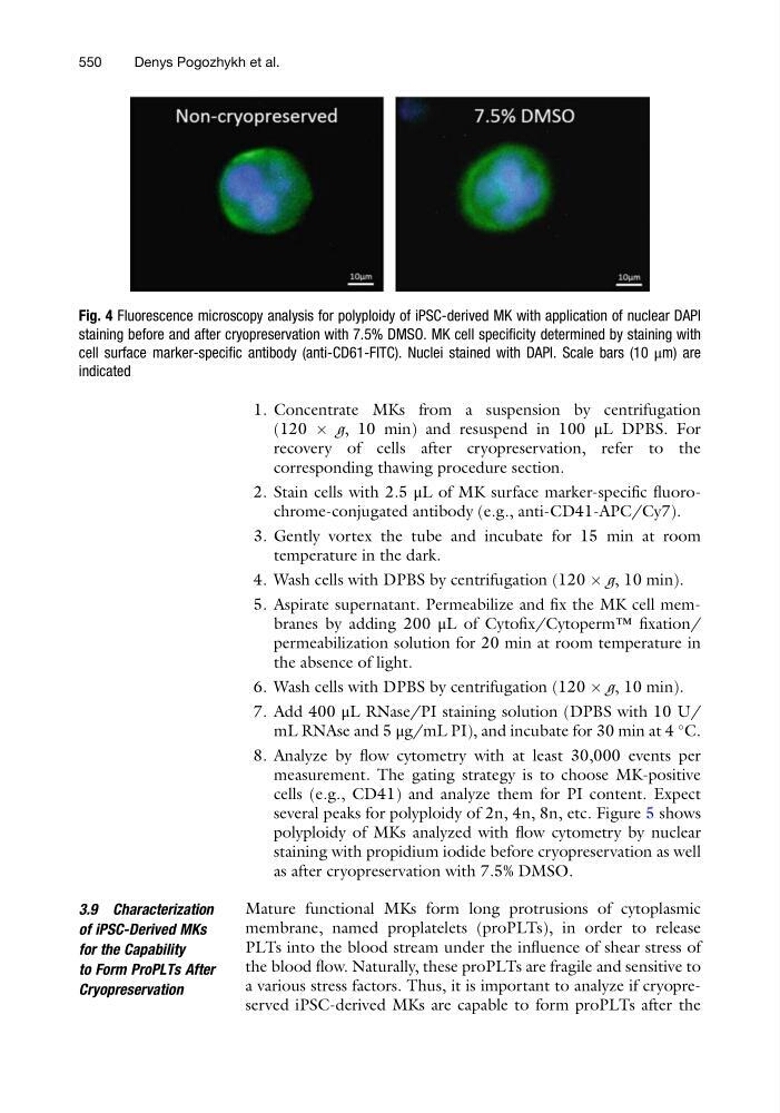
Fig. 4 Fluorescence microscopy analysis for polyploidy of iPSC-derived MK with application of nuclear DAPI
staining before and after cryopreservation with 7.5% DMSO. MK cell specificity determined by staining with
cell surface marker-specific antibody (anti-CD61-FITC). Nuclei stained with DAPI. Scale bars (10 μm) are
indicated
550
Denys Pogozhykh et al.
(120 Â g, 10 min) and resuspend in 100 μL DPBS. For
recovery of cells after cryopreservation, refer to the
corresponding thawing procedure section.
2. Stain cells with 2.5 μL of MK surface marker-specific fluorochrome-conjugated antibody (e.g., anti-CD41-APC/Cy7).
3. Gently vortex the tube and incubate for 15 min at room
temperature in the dark.
4. Wash cells with DPBS by centrifugation (120 Â g, 10 min).
5. Aspirate supernatant. Permeabilize and fix the MK cell membranes by adding 200 μL of Cytofix/Cytoperm™ fixation/
permeabilization solution for 20 min at room temperature in
the absence of light.
6. Wash cells with DPBS by centrifugation (120 Â g, 10 min).
7. Add 400 μL RNase/PI staining solution (DPBS with 10 U/
mL RNAse and 5 μg/mL PI), and incubate for 30 min at 4
C.
8. Analyze by flow cytometry with at least 30,000 events per
measurement. The gating strategy is to choose MK-positive
cells (e.g., CD41) and analyze them for PI content. Expect
several peaks for polyploidy of 2n, 4n, 8n, etc. Figure 5 shows
polyploidy of MKs analyzed with flow cytometry by nuclear
staining with propidium iodide before cryopreservation as well
as after cryopreservation with 7.5% DMSO.
3.9 Characterization
of iPSC-Derived MKs
for the Capability
to Form ProPLTs After
Cryopreservation
Mature functional MKs form long protrusions of cytoplasmic
membrane, named proplatelets (proPLTs), in order to release
PLTs into the blood stream under the influence of shear stress of
the blood flow. Naturally, these proPLTs are fragile and sensitive to
a various stress factors. Thus, it is important to analyze if cryopreserved iPSC-derived MKs are capable to form proPLTs after the
Fig. 4 Fluorescence microscopy analysis for polyploidy of iPSC-derived MK with application of nuclear DAPI
staining before and after cryopreservation with 7.5% DMSO. MK cell specificity determined by staining with
cell surface marker-specific antibody (anti-CD61-FITC). Nuclei stained with DAPI. Scale bars (10 μm) are
indicated
550
Denys Pogozhykh et al.