

3. Transfer 100 μL of cell suspension in a 5 mL test tube.
4. Stain cells with 5 μL of each MK marker-specific fluorochromeconjugated antibody from the panel (e.g., CD41, CD61, and
CD42a). Make sure that fluorochrome spectra of the antibodies do not overlap with each other if measured in a single tube.
Use isotype control antibodies to eliminate unspecific binding.
5. Gently vortex the tube and incubate for 15 min at room
temperature in the dark.
6. Add 200 μL of FACS staining buffer to each tube.
7. Analyze by flow cytometry with at least 10,000 events per
measurement. First, gate the cells according to forward
scatter vs. side scatter (FSC vs. SSC) parameters, and then
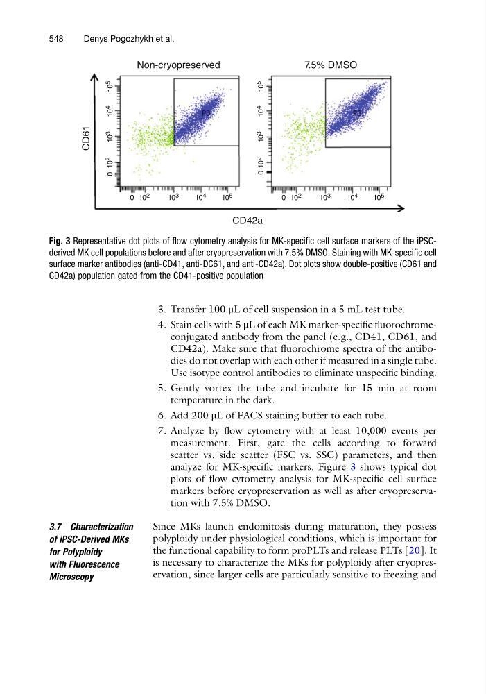
analyze for MK-specific markers. Figure 3 shows typical dot
plots of flow cytometry analysis for MK-specific cell surface
markers before cryopreservation as well as after cryopreservation with 7.5% DMSO.
3.7 Characterization
of iPSC-Derived MKs
for Polyploidy
with Fluorescence
Microscopy
Since MKs launch endomitosis during maturation, they possess
polyploidy under physiological conditions, which is important for
the functional capability to form proPLTs and release PLTs [20]. It
is necessary to characterize the MKs for polyploidy after cryopreservation, since larger cells are particularly sensitive to freezing and
Non-cryopreserved
CD42a
CD61
7.5% DMSO
0 10 2
10 3
10 4
10 5
0 10 2
10 3
10 4
10 5
0 10 2
10 3
10 4
10 5
0 10 2
10 3
10 4
10 5
Fig. 3 Representative dot plots of flow cytometry analysis for MK-specific cell surface markers of the iPSCderived MK cell populations before and after cryopreservation with 7.5% DMSO. Staining with MK-specific cell
surface marker antibodies (anti-CD41, anti-DC61, and anti-CD42a). Dot plots show double-positive (CD61 and
CD42a) population gated from the CD41-positive population
548
Denys Pogozhykh et al.
4. Stain cells with 5 μL of each MK marker-specific fluorochromeconjugated antibody from the panel (e.g., CD41, CD61, and
CD42a). Make sure that fluorochrome spectra of the antibodies do not overlap with each other if measured in a single tube.
Use isotype control antibodies to eliminate unspecific binding.
5. Gently vortex the tube and incubate for 15 min at room
temperature in the dark.
6. Add 200 μL of FACS staining buffer to each tube.
7. Analyze by flow cytometry with at least 10,000 events per
measurement. First, gate the cells according to forward
scatter vs. side scatter (FSC vs. SSC) parameters, and then
analyze for MK-specific markers. Figure 3 shows typical dot
plots of flow cytometry analysis for MK-specific cell surface
markers before cryopreservation as well as after cryopreservation with 7.5% DMSO.
3.7 Characterization
of iPSC-Derived MKs
for Polyploidy
with Fluorescence
Microscopy
Since MKs launch endomitosis during maturation, they possess
polyploidy under physiological conditions, which is important for
the functional capability to form proPLTs and release PLTs [20]. It
is necessary to characterize the MKs for polyploidy after cryopreservation, since larger cells are particularly sensitive to freezing and
Non-cryopreserved
CD42a
CD61
7.5% DMSO
0 10 2
10 3
10 4
10 5
0 10 2
10 3
10 4
10 5
0 10 2
10 3
10 4
10 5
0 10 2
10 3
10 4
10 5
Fig. 3 Representative dot plots of flow cytometry analysis for MK-specific cell surface markers of the iPSCderived MK cell populations before and after cryopreservation with 7.5% DMSO. Staining with MK-specific cell
surface marker antibodies (anti-CD41, anti-DC61, and anti-CD42a). Dot plots show double-positive (CD61 and
CD42a) population gated from the CD41-positive population
548
Denys Pogozhykh et al.