

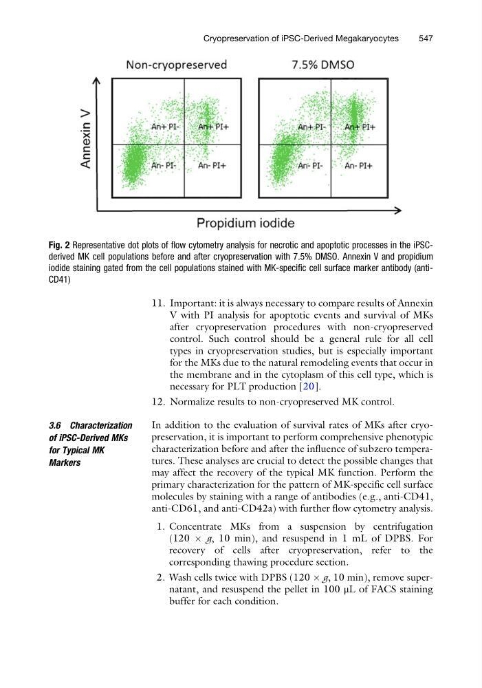
11. Important: it is always necessary to compare results of Annexin
V with PI analysis for apoptotic events and survival of MKs
after cryopreservation procedures with non-cryopreserved
control. Such control should be a general rule for all cell
types in cryopreservation studies, but is especially important
for the MKs due to the natural remodeling events that occur in
the membrane and in the cytoplasm of this cell type, which is
necessary for PLT production [20].
12. Normalize results to non-cryopreserved MK control.
3.6 Characterization
of iPSC-Derived MKs
for Typical MK
Markers
In addition to the evaluation of survival rates of MKs after cryopreservation, it is important to perform comprehensive phenotypic
characterization before and after the influence of subzero temperatures. These analyses are crucial to detect the possible changes that
may affect the recovery of the typical MK function. Perform the
primary characterization for the pattern of MK-specific cell surface
molecules by staining with a range of antibodies (e.g., anti-CD41,
anti-CD61, and anti-CD42a) with further flow cytometry analysis.
1. Concentrate MKs from a suspension by centrifugation
(120 Â g, 10 min), and resuspend in 1 mL of DPBS. For
recovery of cells after cryopreservation, refer to the
corresponding thawing procedure section.
2. Wash cells twice with DPBS (120 Â g, 10 min), remove supernatant, and resuspend the pellet in 100 μL of FACS staining
buffer for each condition.
Fig. 2 Representative dot plots of flow cytometry analysis for necrotic and apoptotic processes in the iPSCderived MK cell populations before and after cryopreservation with 7.5% DMSO. Annexin V and propidium
iodide staining gated from the cell populations stained with MK-specific cell surface marker antibody (antiCD41)
Cryopreservation of iPSC-Derived Megakaryocytes
547
V with PI analysis for apoptotic events and survival of MKs
after cryopreservation procedures with non-cryopreserved
control. Such control should be a general rule for all cell
types in cryopreservation studies, but is especially important
for the MKs due to the natural remodeling events that occur in
the membrane and in the cytoplasm of this cell type, which is
necessary for PLT production [20].
12. Normalize results to non-cryopreserved MK control.
3.6 Characterization
of iPSC-Derived MKs
for Typical MK
Markers
In addition to the evaluation of survival rates of MKs after cryopreservation, it is important to perform comprehensive phenotypic
characterization before and after the influence of subzero temperatures. These analyses are crucial to detect the possible changes that
may affect the recovery of the typical MK function. Perform the
primary characterization for the pattern of MK-specific cell surface
molecules by staining with a range of antibodies (e.g., anti-CD41,
anti-CD61, and anti-CD42a) with further flow cytometry analysis.
1. Concentrate MKs from a suspension by centrifugation
(120 Â g, 10 min), and resuspend in 1 mL of DPBS. For
recovery of cells after cryopreservation, refer to the
corresponding thawing procedure section.
2. Wash cells twice with DPBS (120 Â g, 10 min), remove supernatant, and resuspend the pellet in 100 μL of FACS staining
buffer for each condition.
Fig. 2 Representative dot plots of flow cytometry analysis for necrotic and apoptotic processes in the iPSCderived MK cell populations before and after cryopreservation with 7.5% DMSO. Annexin V and propidium
iodide staining gated from the cell populations stained with MK-specific cell surface marker antibody (antiCD41)
Cryopreservation of iPSC-Derived Megakaryocytes
547