

3 Methods
3.1 Embryo Collapse
1. Prepare a Petri dish with 40 μL droplets of handling medium,
cover with mineral oil, and allow the dish to reach room
temperature (22–25
C). Transfer the expanded blastocyst
stage embryos in these droplets.
2. Place the Petri dish with the embryo on the inverted microscope equipped with a micromanipulation and aspiration system. The collapsing of the embryo should be performed at
room temperature (22–25
C).
3. Hold the embryo in place by suction of the holding pipette
while breaching the capsule or zona pellucida using the aspiration micropipette.
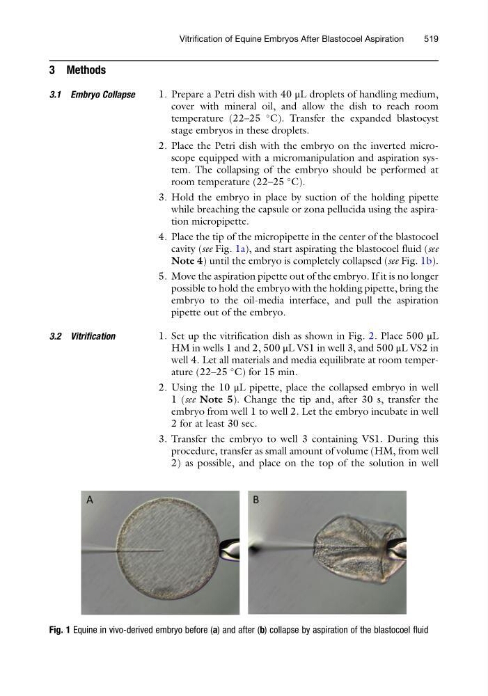
4. Place the tip of the micropipette in the center of the blastocoel
cavity (see Fig. 1a), and start aspirating the blastocoel fluid (see
Note 4) until the embryo is completely collapsed (see Fig. 1b).
5. Move the aspiration pipette out of the embryo. If it is no longer
possible to hold the embryo with the holding pipette, bring the
embryo to the oil-media interface, and pull the aspiration
pipette out of the embryo.
3.2 Vitrification
1. Set up the vitrification dish as shown in Fig. 2. Place 500 μL
HM in wells 1 and 2, 500 μL VS1 in well 3, and 500 μL VS2 in
well 4. Let all materials and media equilibrate at room temperature (22–25
C) for 15 min.
2. Using the 10 μL pipette, place the collapsed embryo in well
1 (see Note 5). Change the tip and, after 30 s, transfer the
embryo from well 1 to well 2. Let the embryo incubate in well
2 for at least 30 sec.
3. Transfer the embryo to well 3 containing VS1. During this
procedure, transfer as small amount of volume (HM, from well
2) as possible, and place on the top of the solution in well
Fig. 1 Equine in vivo-derived embryo before (a) and after (b) collapse by aspiration of the blastocoel fluid
Vitrification of Equine Embryos After Blastocoel Aspiration
519
3.1 Embryo Collapse
1. Prepare a Petri dish with 40 μL droplets of handling medium,
cover with mineral oil, and allow the dish to reach room
temperature (22–25
C). Transfer the expanded blastocyst
stage embryos in these droplets.
2. Place the Petri dish with the embryo on the inverted microscope equipped with a micromanipulation and aspiration system. The collapsing of the embryo should be performed at
room temperature (22–25
C).
3. Hold the embryo in place by suction of the holding pipette
while breaching the capsule or zona pellucida using the aspiration micropipette.
4. Place the tip of the micropipette in the center of the blastocoel
cavity (see Fig. 1a), and start aspirating the blastocoel fluid (see
Note 4) until the embryo is completely collapsed (see Fig. 1b).
5. Move the aspiration pipette out of the embryo. If it is no longer
possible to hold the embryo with the holding pipette, bring the
embryo to the oil-media interface, and pull the aspiration
pipette out of the embryo.
3.2 Vitrification
1. Set up the vitrification dish as shown in Fig. 2. Place 500 μL
HM in wells 1 and 2, 500 μL VS1 in well 3, and 500 μL VS2 in
well 4. Let all materials and media equilibrate at room temperature (22–25
C) for 15 min.
2. Using the 10 μL pipette, place the collapsed embryo in well
1 (see Note 5). Change the tip and, after 30 s, transfer the
embryo from well 1 to well 2. Let the embryo incubate in well
2 for at least 30 sec.
3. Transfer the embryo to well 3 containing VS1. During this
procedure, transfer as small amount of volume (HM, from well
2) as possible, and place on the top of the solution in well
Fig. 1 Equine in vivo-derived embryo before (a) and after (b) collapse by aspiration of the blastocoel fluid
Vitrification of Equine Embryos After Blastocoel Aspiration
519