

into a Petri dish containing 3–4 mL room temperature
TL-HEPES solution.
3. Carefully dissect out the fat pad, ovarian bursa, and oviduct
tissues under a stereo dissecting microscope using a fine forceps
and micro-scissors.
4. Transfer the cleaned ovaries into a Petri dish containing fresh
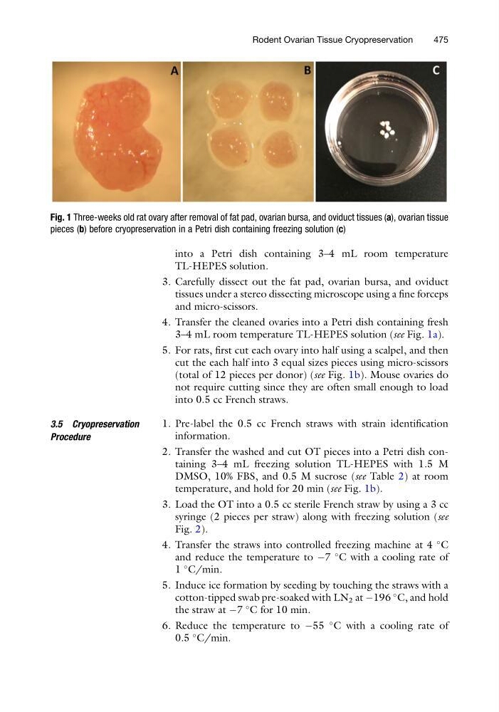
3–4 mL room temperature TL-HEPES solution (see Fig. 1a).
5. For rats, first cut each ovary into half using a scalpel, and then
cut the each half into 3 equal sizes pieces using micro-scissors
(total of 12 pieces per donor) (see Fig. 1b). Mouse ovaries do
not require cutting since they are often small enough to load
into 0.5 cc French straws.
3.5 Cryopreservation
Procedure
1. Pre-label the 0.5 cc French straws with strain identification
information.
2. Transfer the washed and cut OT pieces into a Petri dish containing 3–4 mL freezing solution TL-HEPES with 1.5 M
DMSO, 10% FBS, and 0.5 M sucrose (see Table 2) at room
temperature, and hold for 20 min (see Fig. 1b).
3. Load the OT into a 0.5 cc sterile French straw by using a 3 cc
syringe (2 pieces per straw) along with freezing solution (see
Fig. 2).
4. Transfer the straws into controlled freezing machine at 4
C
and reduce the temperature to À7
C with a cooling rate of
1
C/min.
5. Induce ice formation by seeding by touching the straws with a
cotton-tipped swab pre-soaked with LN 2 at À196
C, and hold
the straw at À7
C for 10 min.
6. Reduce the temperature to À55
C with a cooling rate of
0.5
C/min.
Fig. 1 Three-weeks old rat ovary after removal of fat pad, ovarian bursa, and oviduct tissues (a), ovarian tissue
pieces (b) before cryopreservation in a Petri dish containing freezing solution (c)
Rodent Ovarian Tissue Cryopreservation
475
TL-HEPES solution.
3. Carefully dissect out the fat pad, ovarian bursa, and oviduct
tissues under a stereo dissecting microscope using a fine forceps
and micro-scissors.
4. Transfer the cleaned ovaries into a Petri dish containing fresh
3–4 mL room temperature TL-HEPES solution (see Fig. 1a).
5. For rats, first cut each ovary into half using a scalpel, and then
cut the each half into 3 equal sizes pieces using micro-scissors
(total of 12 pieces per donor) (see Fig. 1b). Mouse ovaries do
not require cutting since they are often small enough to load
into 0.5 cc French straws.
3.5 Cryopreservation
Procedure
1. Pre-label the 0.5 cc French straws with strain identification
information.
2. Transfer the washed and cut OT pieces into a Petri dish containing 3–4 mL freezing solution TL-HEPES with 1.5 M
DMSO, 10% FBS, and 0.5 M sucrose (see Table 2) at room
temperature, and hold for 20 min (see Fig. 1b).
3. Load the OT into a 0.5 cc sterile French straw by using a 3 cc
syringe (2 pieces per straw) along with freezing solution (see
Fig. 2).
4. Transfer the straws into controlled freezing machine at 4
C
and reduce the temperature to À7
C with a cooling rate of
1
C/min.
5. Induce ice formation by seeding by touching the straws with a
cotton-tipped swab pre-soaked with LN 2 at À196
C, and hold
the straw at À7
C for 10 min.
6. Reduce the temperature to À55
C with a cooling rate of
0.5
C/min.
Fig. 1 Three-weeks old rat ovary after removal of fat pad, ovarian bursa, and oviduct tissues (a), ovarian tissue
pieces (b) before cryopreservation in a Petri dish containing freezing solution (c)
Rodent Ovarian Tissue Cryopreservation
475