

3.4 Acquisition of
Transmission Spectra
Using the
TemperatureControlled Sample
Holder
1. Concentrate the sample by centrifugation (e.g., 0.5–1 mL cell
or lipid suspension, 1200 Â g for 1 min) and transfer the
obtained pellet (i.e., hydrated sample) on a CaF 2 window
(e.g., 20 μL cell pellet or 2–5 μL concentrated lipid sample).
Add a second window such that the sample is sandwiched
between two windows. For dried specimens, use a Teflon
spacer between the windows to avoid sticking at high temperatures. Grease can be used around the windows to seal the
sample and prevent dehydration during heating.
2. Place the windows with the sample in the temperaturecontrolled sample holder and insert the thermocouple such
that it touches the edge of the sample (via a Luer lock, grease
can be used to facilitate contact). Check connections for heating and cooling of the sample holder, close the lid around the
sample holder area, and wait about 5–10 min until the area is
thoroughly flushed with dry air from the purge system.
3. Acquire a spectrum of the sample using appropriate settings
(8 co-added interferograms, 4000–900 cm
À1 wave number
range, 4 cm
À1 resolution, in absorbance), to verify the quality
of the spectrum. Prepare a new sample if needed (see Note 14).
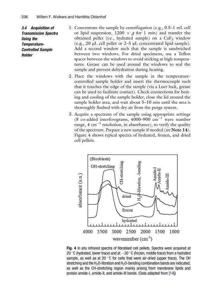
Figure 4 shows typical spectra of hydrated, frozen, and dried
cell pellets.
Fig. 4 In situ infrared spectra of fibroblast cell pellets. Spectra were acquired at
20
C (hydrated, lower trace) and at À30
C (frozen, middle trace) from a hydrated
sample, as well as at 20
C for cells that were air-dried (upper trace). The OH
stretching and the H 2 O-libration and H 2 O-bending combination bands are indicated,
as well as the CH-stretching region mainly arising from membrane lipids and
protein amide-I, amide-II, and amide-III bands. (Data adapted from [14])
338
Willem F. Wolkers and Harrie ¨ tte Oldenhof
Transmission Spectra
Using the
TemperatureControlled Sample
Holder
1. Concentrate the sample by centrifugation (e.g., 0.5–1 mL cell
or lipid suspension, 1200 Â g for 1 min) and transfer the
obtained pellet (i.e., hydrated sample) on a CaF 2 window
(e.g., 20 μL cell pellet or 2–5 μL concentrated lipid sample).
Add a second window such that the sample is sandwiched
between two windows. For dried specimens, use a Teflon
spacer between the windows to avoid sticking at high temperatures. Grease can be used around the windows to seal the
sample and prevent dehydration during heating.
2. Place the windows with the sample in the temperaturecontrolled sample holder and insert the thermocouple such
that it touches the edge of the sample (via a Luer lock, grease
can be used to facilitate contact). Check connections for heating and cooling of the sample holder, close the lid around the
sample holder area, and wait about 5–10 min until the area is
thoroughly flushed with dry air from the purge system.
3. Acquire a spectrum of the sample using appropriate settings
(8 co-added interferograms, 4000–900 cm
À1 wave number
range, 4 cm
À1 resolution, in absorbance), to verify the quality
of the spectrum. Prepare a new sample if needed (see Note 14).
Figure 4 shows typical spectra of hydrated, frozen, and dried
cell pellets.
Fig. 4 In situ infrared spectra of fibroblast cell pellets. Spectra were acquired at
20
C (hydrated, lower trace) and at À30
C (frozen, middle trace) from a hydrated
sample, as well as at 20
C for cells that were air-dried (upper trace). The OH
stretching and the H 2 O-libration and H 2 O-bending combination bands are indicated,
as well as the CH-stretching region mainly arising from membrane lipids and
protein amide-I, amide-II, and amide-III bands. (Data adapted from [14])
338
Willem F. Wolkers and Harrie ¨ tte Oldenhof