

and reflection mode in case of the ATR accessory; and (5) select
the detector (MCT or TGS) and adjust the iris opening if
needed (see Note 10).
5. Acquire a background spectrum and save it. Make sure that the
sample area is thoroughly flushed with dry air and that the
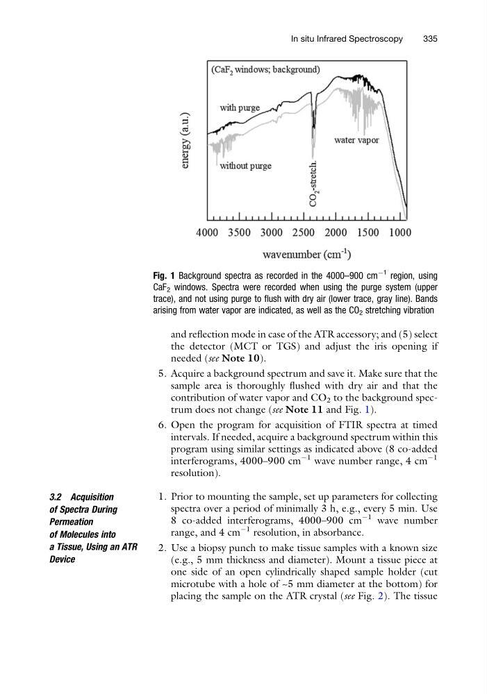
contribution of water vapor and CO 2 to the background spectrum does not change (see Note 11 and Fig. 1).
6. Open the program for acquisition of FTIR spectra at timed
intervals. If needed, acquire a background spectrum within this
program using similar settings as indicated above (8 co-added
interferograms, 4000–900 cm
À1 wave number range, 4 cm
À1
resolution).
3.2 Acquisition
of Spectra During
Permeation
of Molecules into
a Tissue, Using an ATR
Device
1. Prior to mounting the sample, set up parameters for collecting
spectra over a period of minimally 3 h, e.g., every 5 min. Use
8 co-added interferograms, 4000–900 cm
À1 wave number
range, and 4 cm
À1 resolution, in absorbance.
2. Use a biopsy punch to make tissue samples with a known size
(e.g., 5 mm thickness and diameter). Mount a tissue piece at
one side of an open cylindrically shaped sample holder (cut
microtube with a hole of ~5 mm diameter at the bottom) for
placing the sample on the ATR crystal (see Fig. 2). The tissue
Fig. 1 Background spectra as recorded in the 4000–900 cm
À1 region, using
CaF 2 windows. Spectra were recorded when using the purge system (upper
trace), and not using purge to flush with dry air (lower trace, gray line). Bands
arising from water vapor are indicated, as well as the CO 2 stretching vibration
In situ Infrared Spectroscopy
335
the detector (MCT or TGS) and adjust the iris opening if
needed (see Note 10).
5. Acquire a background spectrum and save it. Make sure that the
sample area is thoroughly flushed with dry air and that the
contribution of water vapor and CO 2 to the background spectrum does not change (see Note 11 and Fig. 1).
6. Open the program for acquisition of FTIR spectra at timed
intervals. If needed, acquire a background spectrum within this
program using similar settings as indicated above (8 co-added
interferograms, 4000–900 cm
À1 wave number range, 4 cm
À1
resolution).
3.2 Acquisition
of Spectra During
Permeation
of Molecules into
a Tissue, Using an ATR
Device
1. Prior to mounting the sample, set up parameters for collecting
spectra over a period of minimally 3 h, e.g., every 5 min. Use
8 co-added interferograms, 4000–900 cm
À1 wave number
range, and 4 cm
À1 resolution, in absorbance.
2. Use a biopsy punch to make tissue samples with a known size
(e.g., 5 mm thickness and diameter). Mount a tissue piece at
one side of an open cylindrically shaped sample holder (cut
microtube with a hole of ~5 mm diameter at the bottom) for
placing the sample on the ATR crystal (see Fig. 2). The tissue
Fig. 1 Background spectra as recorded in the 4000–900 cm
À1 region, using
CaF 2 windows. Spectra were recorded when using the purge system (upper
trace), and not using purge to flush with dry air (lower trace, gray line). Bands
arising from water vapor are indicated, as well as the CO 2 stretching vibration
In situ Infrared Spectroscopy
335