

by pumping dry air into a chamber containing the experimental
setup as shown in Fig. 6 and monitoring the RH with a temperature
and RH logger (e.g., ONSET UX100-011).
3.2.1 Laser-Assisted
Drying and Storage
of Proteins
Drying methods are described for egg white lysozyme (Worthington Biochemical) as example.
1. Mixing with trehalose: Prepare a drying solution
(DS) consisting of 0.2 M trehalose in 0.33Â phosphate buffer
solution (PBS) (see Note 7). Prepare 40 μL droplets containing
egg white lysozyme dissolved in DS at a concentration of
0.5 mg/mL (see Note 8).
2. Loading samples onto coverslips: Deposit a 40 μL droplet of
the protein-drying solution onto an 18 mm diameter borosilicate glass coverslip (Fisherbrand).
3. Determine the initial mass gravimetrically using a microbalance. Mark a fiducial on the edge of the coverslip to ensure
consistent orientation for imaging conducted during the study.
4. Laser-assisted drying: Move the sample into the low humidity
chamber (23
C, 11% RH) for laser irradiation. Process samples
for 60 min at 5 W (26.9 W/cm
2 ). Monitor the maximum
temperature of the sample during processing using the thermal
camera.
5. End moisture content determination: Remove the sample from
the humidity chamber, and immediately determine the mass
again. The end moisture content (EMC), which is a measure of
the amount of water relative to the dry mass of a sample, can be
calculated as:
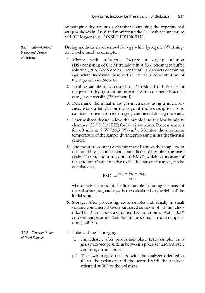
EMC ¼
m f À m s À m dw
m dw
where m f is the mass of the final sample including the mass of
the substrate, m s , and m dw is the calculated dry weight of the
initial sample.
6. Storage: After processing, store samples individually in small
volume containers above a saturated solution of lithium chloride. The RH of above a saturated LiCl solution is 14.3 Æ 0.5%
at room temperature. Samples can be stored at room temperature (~23
C).
3.2.2 Characterization
of Dried Samples
1. Polarized Light Imaging.
(a) Immediately after processing, place LAD samples on a
glass microscope slide in between a polarizer and analyzer,
and image from above.
(b) Take two images: the first with the analyzer oriented at
0
to the polarizer and the second with the analyzer
oriented at 90
to the polarizer.
Drying Technology for Preservation of Biologics
217
setup as shown in Fig. 6 and monitoring the RH with a temperature
and RH logger (e.g., ONSET UX100-011).
3.2.1 Laser-Assisted
Drying and Storage
of Proteins
Drying methods are described for egg white lysozyme (Worthington Biochemical) as example.
1. Mixing with trehalose: Prepare a drying solution
(DS) consisting of 0.2 M trehalose in 0.33Â phosphate buffer
solution (PBS) (see Note 7). Prepare 40 μL droplets containing
egg white lysozyme dissolved in DS at a concentration of
0.5 mg/mL (see Note 8).
2. Loading samples onto coverslips: Deposit a 40 μL droplet of
the protein-drying solution onto an 18 mm diameter borosilicate glass coverslip (Fisherbrand).
3. Determine the initial mass gravimetrically using a microbalance. Mark a fiducial on the edge of the coverslip to ensure
consistent orientation for imaging conducted during the study.
4. Laser-assisted drying: Move the sample into the low humidity
chamber (23
C, 11% RH) for laser irradiation. Process samples
for 60 min at 5 W (26.9 W/cm
2 ). Monitor the maximum
temperature of the sample during processing using the thermal
camera.
5. End moisture content determination: Remove the sample from
the humidity chamber, and immediately determine the mass
again. The end moisture content (EMC), which is a measure of
the amount of water relative to the dry mass of a sample, can be
calculated as:
EMC ¼
m f À m s À m dw
m dw
where m f is the mass of the final sample including the mass of
the substrate, m s , and m dw is the calculated dry weight of the
initial sample.
6. Storage: After processing, store samples individually in small
volume containers above a saturated solution of lithium chloride. The RH of above a saturated LiCl solution is 14.3 Æ 0.5%
at room temperature. Samples can be stored at room temperature (~23
C).
3.2.2 Characterization
of Dried Samples
1. Polarized Light Imaging.
(a) Immediately after processing, place LAD samples on a
glass microscope slide in between a polarizer and analyzer,
and image from above.
(b) Take two images: the first with the analyzer oriented at
0
to the polarizer and the second with the analyzer
oriented at 90
to the polarizer.
Drying Technology for Preservation of Biologics
217