

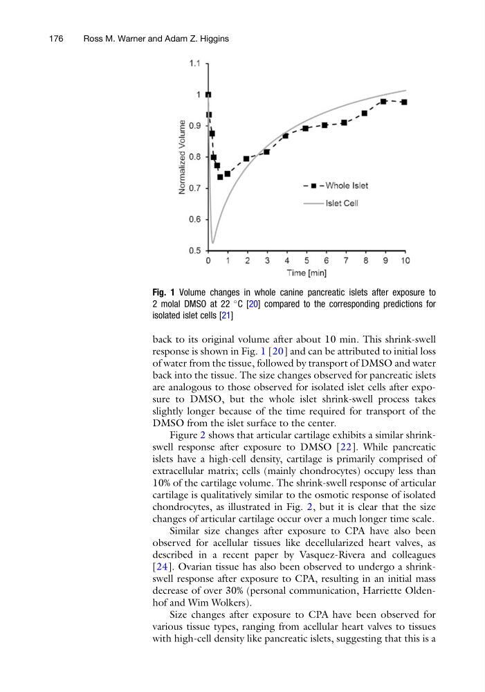
back to its original volume after about 10 min. This shrink-swell
response is shown in Fig. 1 [20] and can be attributed to initial loss
of water from the tissue, followed by transport of DMSO and water
back into the tissue. The size changes observed for pancreatic islets
are analogous to those observed for isolated islet cells after exposure to DMSO, but the whole islet shrink-swell process takes
slightly longer because of the time required for transport of the
DMSO from the islet surface to the center.
Figure 2 shows that articular cartilage exhibits a similar shrinkswell response after exposure to DMSO [22]. While pancreatic
islets have a high-cell density, cartilage is primarily comprised of
extracellular matrix; cells (mainly chondrocytes) occupy less than
10% of the cartilage volume. The shrink-swell response of articular
cartilage is qualitatively similar to the osmotic response of isolated
chondrocytes, as illustrated in Fig. 2, but it is clear that the size
changes of articular cartilage occur over a much longer time scale.
Similar size changes after exposure to CPA have also been
observed for acellular tissues like decellularized heart valves, as
described in a recent paper by Vasquez-Rivera and colleagues
[24]. Ovarian tissue has also been observed to undergo a shrinkswell response after exposure to CPA, resulting in an initial mass
decrease of over 30% (personal communication, Harriette Oldenhof and Wim Wolkers).
Size changes after exposure to CPA have been observed for
various tissue types, ranging from acellular heart valves to tissues
with high-cell density like pancreatic islets, suggesting that this is a
Fig. 1 Volume changes in whole canine pancreatic islets after exposure to
2 molal DMSO at 22
C [20] compared to the corresponding predictions for
isolated islet cells [21]
176
Ross M. Warner and Adam Z. Higgins
response is shown in Fig. 1 [20] and can be attributed to initial loss
of water from the tissue, followed by transport of DMSO and water
back into the tissue. The size changes observed for pancreatic islets
are analogous to those observed for isolated islet cells after exposure to DMSO, but the whole islet shrink-swell process takes
slightly longer because of the time required for transport of the
DMSO from the islet surface to the center.
Figure 2 shows that articular cartilage exhibits a similar shrinkswell response after exposure to DMSO [22]. While pancreatic
islets have a high-cell density, cartilage is primarily comprised of
extracellular matrix; cells (mainly chondrocytes) occupy less than
10% of the cartilage volume. The shrink-swell response of articular
cartilage is qualitatively similar to the osmotic response of isolated
chondrocytes, as illustrated in Fig. 2, but it is clear that the size
changes of articular cartilage occur over a much longer time scale.
Similar size changes after exposure to CPA have also been
observed for acellular tissues like decellularized heart valves, as
described in a recent paper by Vasquez-Rivera and colleagues
[24]. Ovarian tissue has also been observed to undergo a shrinkswell response after exposure to CPA, resulting in an initial mass
decrease of over 30% (personal communication, Harriette Oldenhof and Wim Wolkers).
Size changes after exposure to CPA have been observed for
various tissue types, ranging from acellular heart valves to tissues
with high-cell density like pancreatic islets, suggesting that this is a
Fig. 1 Volume changes in whole canine pancreatic islets after exposure to
2 molal DMSO at 22
C [20] compared to the corresponding predictions for
isolated islet cells [21]
176
Ross M. Warner and Adam Z. Higgins