

–1
–1
–1
–1
8
–1
–1
–1
–1
70
Biomedical Signal and Image Processing
As described earlier, the image segmentation techniques can be classified into
two general categories. In the first category of techniques, segmentation is conducted based on the discontinuity of the points across two regions, while, in the
second group of segmentation methods, the algorithms exploit the similarities
among the points in the same region for segmentation. We first focus on the first
category.
The main methods in the first category include detecting gray-level discontinuities such as points, lines, and edges. Another popular method in the first category
is thresholding. In thresholding, a part of the image is selected based on its graylevel difference from the other parts of the image. Here, we first introduce some
methods for detection of points and lines in an image. Then, we will describe
image segmentation methods that detect regions and objects in an image using
thresholding ideas.
4.3.1 POINT DETECTION
In point detection methods, the intention is to detect the isolated points in an
image. The main factor that can help us detect these isolated points or pixels is the
difference between their gray levels and gray levels of their neighboring pixels.
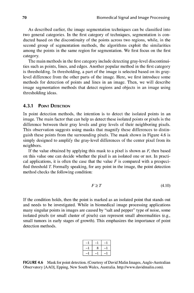
This observation suggests using masks that magnify these differences to distinguish these points from the surrounding pixels. The mask shown in Figure 4.6 is
simply designed to amplify the gray-level differences of the center pixel from its
neighbors.
If the value obtained by applying this mask to a pixel is shown as F, then based
on this value one can decide whether the pixel is an isolated one or not. In practical applications, it is often the case that the value F is compared with a prespecified threshold T. Formally speaking, for any point in the image, the point detection
method checks the following condition:
F T
(4.10)
≥
If the condition holds, then the point is marked as an isolated point that stands out
and needs to be investigated. While in biomedical image processing applications
many singular points in images are caused by “salt and pepper” type of noise, some
isolated pixels (or small cluster of pixels) can represent small abnormalities (e.g.,
small tumors in early stages of growth). This emphasizes the importance of point
detection methods.
FIGURE 4.6 Mask for point detection. (Courtesy of David Malin Images, Anglo-Australian
Observatory [AAO], Epping, New South Wales, Australia. http://www.davidmalin.com).
–1
–1
–1
8
–1
–1
–1
–1
70
Biomedical Signal and Image Processing
As described earlier, the image segmentation techniques can be classified into
two general categories. In the first category of techniques, segmentation is conducted based on the discontinuity of the points across two regions, while, in the
second group of segmentation methods, the algorithms exploit the similarities
among the points in the same region for segmentation. We first focus on the first
category.
The main methods in the first category include detecting gray-level discontinuities such as points, lines, and edges. Another popular method in the first category
is thresholding. In thresholding, a part of the image is selected based on its graylevel difference from the other parts of the image. Here, we first introduce some
methods for detection of points and lines in an image. Then, we will describe
image segmentation methods that detect regions and objects in an image using
thresholding ideas.
4.3.1 POINT DETECTION
In point detection methods, the intention is to detect the isolated points in an
image. The main factor that can help us detect these isolated points or pixels is the
difference between their gray levels and gray levels of their neighboring pixels.
This observation suggests using masks that magnify these differences to distinguish these points from the surrounding pixels. The mask shown in Figure 4.6 is
simply designed to amplify the gray-level differences of the center pixel from its
neighbors.
If the value obtained by applying this mask to a pixel is shown as F, then based
on this value one can decide whether the pixel is an isolated one or not. In practical applications, it is often the case that the value F is compared with a prespecified threshold T. Formally speaking, for any point in the image, the point detection
method checks the following condition:
F T
(4.10)
≥
If the condition holds, then the point is marked as an isolated point that stands out
and needs to be investigated. While in biomedical image processing applications
many singular points in images are caused by “salt and pepper” type of noise, some
isolated pixels (or small cluster of pixels) can represent small abnormalities (e.g.,
small tumors in early stages of growth). This emphasizes the importance of point
detection methods.
FIGURE 4.6 Mask for point detection. (Courtesy of David Malin Images, Anglo-Australian
Observatory [AAO], Epping, New South Wales, Australia. http://www.davidmalin.com).