

4
Edge Detection and
Segmentation of Images
4.1 INTRODUCTION AND OVERVIEW
In medical image processing, as well as many other applications of image processing, it is necessary to identify the boundary between the objects in the image and
separate the objects from each other. For example, when analyzing a cell image
(captured by a microscope), it is vital to design image processing algorithms to segment the image and distinguish the objects in the cell from each other, for example,
identify the contour of a nucleus. In many practical applications, segmentation and
edge detection techniques allow separation of objects residing in an image and identify the boundary among them.
In principle, there are two approaches for edge detection and segmentation. In the
first approach, differences and dissimilarities of pixels in two neighboring regions
(objects) are exploited to segment the two regions, while, in the second approach, the
similarities of the pixels within each region are used to separate the region from the
neighboring regions. As can be seen, while the two approaches are based on rather
related ideas, they apply different criteria. We discuss several examples of each of
these two approaches in this chapter.
4.2 EDGE DETECTION
In this section, some of the main edge detection techniques, commonly used in biomedical image processing, are reviewed and compared with each other.
4.2.1 SOBEL EDGE DETECTION
The Sobel technique is one of the most popular edge detection techniques that is also
computationally simple. In this technique, a 3 × 3 simple mask is used to magnify
the differences among the points on the opposite sides of a boundary and eliminate
the smooth gray-level changes in the pixels located on the same side of the boundary.
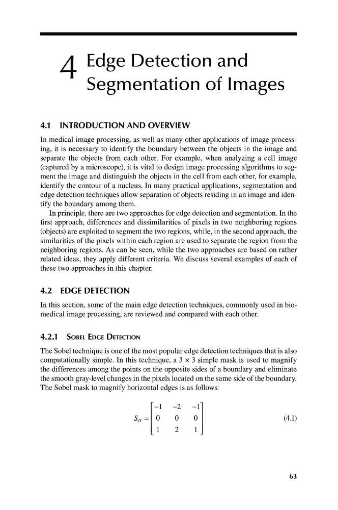
The Sobel mask to magnify horizontal edges is as follows:
⎡
−
−2
1
0
0
0
−
1
⎤
⎥
⎥
⎥ ⎦
⎢
⎢
⎢ ⎣
S H =
(4.1)
1
2
1
63
Edge Detection and
Segmentation of Images
4.1 INTRODUCTION AND OVERVIEW
In medical image processing, as well as many other applications of image processing, it is necessary to identify the boundary between the objects in the image and
separate the objects from each other. For example, when analyzing a cell image
(captured by a microscope), it is vital to design image processing algorithms to segment the image and distinguish the objects in the cell from each other, for example,
identify the contour of a nucleus. In many practical applications, segmentation and
edge detection techniques allow separation of objects residing in an image and identify the boundary among them.
In principle, there are two approaches for edge detection and segmentation. In the
first approach, differences and dissimilarities of pixels in two neighboring regions
(objects) are exploited to segment the two regions, while, in the second approach, the
similarities of the pixels within each region are used to separate the region from the
neighboring regions. As can be seen, while the two approaches are based on rather
related ideas, they apply different criteria. We discuss several examples of each of
these two approaches in this chapter.
4.2 EDGE DETECTION
In this section, some of the main edge detection techniques, commonly used in biomedical image processing, are reviewed and compared with each other.
4.2.1 SOBEL EDGE DETECTION
The Sobel technique is one of the most popular edge detection techniques that is also
computationally simple. In this technique, a 3 × 3 simple mask is used to magnify
the differences among the points on the opposite sides of a boundary and eliminate
the smooth gray-level changes in the pixels located on the same side of the boundary.
The Sobel mask to magnify horizontal edges is as follows:
⎡
−
−2
1
0
0
0
−
1
⎤
⎥
⎥
⎥ ⎦
⎢
⎢
⎢ ⎣
S H =
(4.1)
1
2
1
63