

374
Biomedical Signal and Image Processing
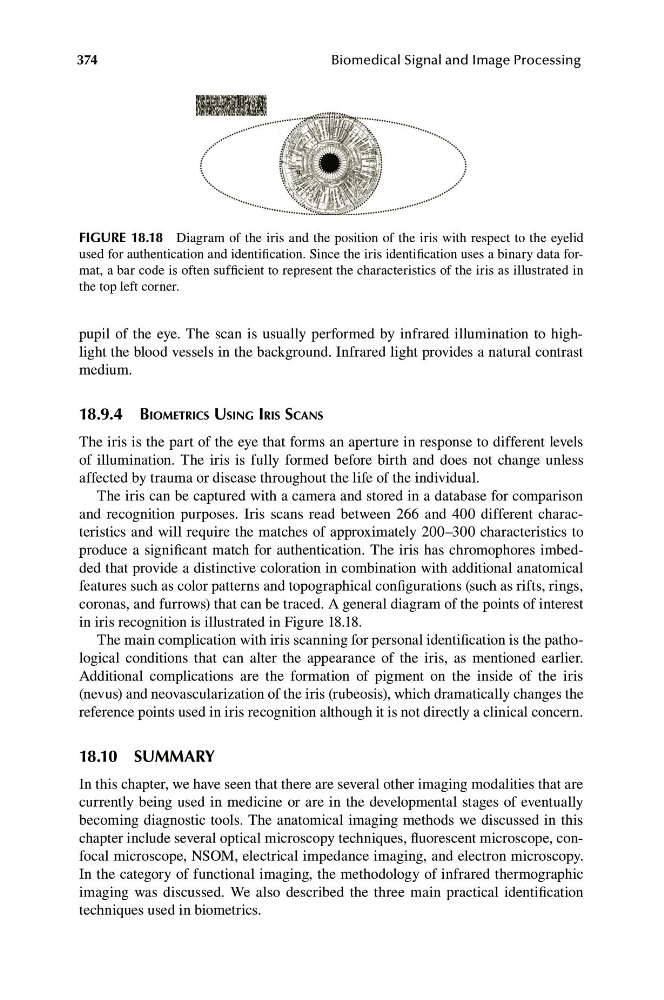
FIGURE 18.18 Diagram of the iris and the position of the iris with respect to the eyelid
used for authentication and identification. Since the iris identification uses a binary data format, a bar code is often sufficient to represent the characteristics of the iris as illustrated in
the top left corner.
pupil of the eye. The scan is usually performed by infrared illumination to highlight the blood vessels in the background. Infrared light provides a natural contrast
medium.
18.9.4 BIOMETRICS USING IRIS SCANS
The iris is the part of the eye that forms an aperture in response to different levels
of illumination. The iris is fully formed before birth and does not change unless
affected by trauma or disease throughout the life of the individual.
The iris can be captured with a camera and stored in a database for comparison
and recognition purposes. Iris scans read between 266 and 400 different characteristics and will require the matches of approximately 200–300 characteristics to
produce a significant match for authentication. The iris has chromophores imbedded that provide a distinctive coloration in combination with additional anatomical
features such as color patterns and topographical configurations (such as rifts, rings,
coronas, and furrows) that can be traced. A general diagram of the points of interest
in iris recognition is illustrated in Figure 18.18.
The main complication with iris scanning for personal identification is the pathological conditions that can alter the appearance of the iris, as mentioned earlier.
Additional complications are the formation of pigment on the inside of the iris
(nevus) and neovascularization of the iris (rubeosis), which dramatically changes the
reference points used in iris recognition although it is not directly a clinical concern.
18.10 SUMMARY
In this chapter, we have seen that there are several other imaging modalities that are
currently being used in medicine or are in the developmental stages of eventually
becoming diagnostic tools. The anatomical imaging methods we discussed in this
chapter include several optical microscopy techniques, fluorescent microscope, confocal microscope, NSOM, electrical impedance imaging, and electron microscopy.
In the category of functional imaging, the methodology of infrared thermographic
imaging was discussed. We also described the three main practical identification
techniques used in biometrics.
Biomedical Signal and Image Processing
FIGURE 18.18 Diagram of the iris and the position of the iris with respect to the eyelid
used for authentication and identification. Since the iris identification uses a binary data format, a bar code is often sufficient to represent the characteristics of the iris as illustrated in
the top left corner.
pupil of the eye. The scan is usually performed by infrared illumination to highlight the blood vessels in the background. Infrared light provides a natural contrast
medium.
18.9.4 BIOMETRICS USING IRIS SCANS
The iris is the part of the eye that forms an aperture in response to different levels
of illumination. The iris is fully formed before birth and does not change unless
affected by trauma or disease throughout the life of the individual.
The iris can be captured with a camera and stored in a database for comparison
and recognition purposes. Iris scans read between 266 and 400 different characteristics and will require the matches of approximately 200–300 characteristics to
produce a significant match for authentication. The iris has chromophores imbedded that provide a distinctive coloration in combination with additional anatomical
features such as color patterns and topographical configurations (such as rifts, rings,
coronas, and furrows) that can be traced. A general diagram of the points of interest
in iris recognition is illustrated in Figure 18.18.
The main complication with iris scanning for personal identification is the pathological conditions that can alter the appearance of the iris, as mentioned earlier.
Additional complications are the formation of pigment on the inside of the iris
(nevus) and neovascularization of the iris (rubeosis), which dramatically changes the
reference points used in iris recognition although it is not directly a clinical concern.
18.10 SUMMARY
In this chapter, we have seen that there are several other imaging modalities that are
currently being used in medicine or are in the developmental stages of eventually
becoming diagnostic tools. The anatomical imaging methods we discussed in this
chapter include several optical microscopy techniques, fluorescent microscope, confocal microscope, NSOM, electrical impedance imaging, and electron microscopy.
In the category of functional imaging, the methodology of infrared thermographic
imaging was discussed. We also described the three main practical identification
techniques used in biometrics.