

373
Other Biomedical Imaging Techniques
called minutiae. The characteristics are organized based on type of formation,
orientation, spatial frequency, curvature, and position of arches. Some of the fingerprint minutiae are crossovers in the ridges, the shape of the core of an arch, islands,
delta-shaped ridge formations, and seemingly bidirectional deadened ridges.
When these features are captures for a given person, they are matched against the
same features captured for all cases in the dataset. Fingerprinting is the most commonly used biometrics method and is heavily used in security applications.
18.9.3 BIOMETRICS USING RETINA SCANS
Retina scans are invaluable tools for personal identification purposes. A retinal scan
analyzes the topographical distribution of blood vessels in the retina, using the fovea
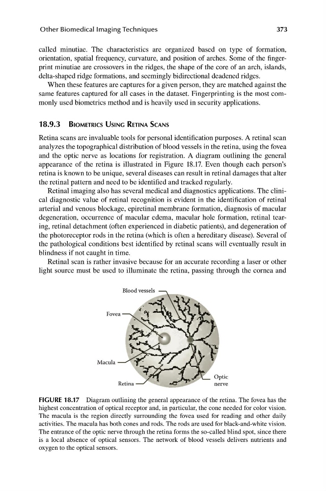
and the optic nerve as locations for registration. A diagram outlining the general
appearance of the retina is illustrated in Figure 18.17. Even though each person’s
retina is known to be unique, several diseases can result in retinal damages that alter
the retinal pattern and need to be identified and tracked regularly.
Retinal imaging also has several medical and diagnostics applications. The clinical diagnostic value of retinal recognition is evident in the identification of retinal
arterial and venous blockage, epiretinal membrane formation, diagnosis of macular
degeneration, occurrence of macular edema, macular hole formation, retinal tearing, retinal detachment (often experienced in diabetic patients), and degeneration of
the photoreceptor rods in the retina (which is often a hereditary disease). Several of
the pathological conditions best identified by retinal scans will eventually result in
blindness if not caught in time.
Retinal scan is rather invasive because for an accurate recording a laser or other
light source must be used to illuminate the retina, passing through the cornea and
Blood vessels
Fovea
Macula
Retina
Optic
nerve
FIGURE 18.17 Diagram outlining the general appearance of the retina. The fovea has the
highest concentration of optical receptor and, in particular, the cone needed for color vision.
The macula is the region directly surrounding the fovea used for reading and other daily
activities. The macula has both cones and rods. The rods are used for black-and-white vision.
The entrance of the optic nerve through the retina forms the so-called blind spot, since there
is a local absence of optical sensors. The network of blood vessels delivers nutrients and
oxygen to the optical sensors.