

353
L
A
Positron Emission Tomography
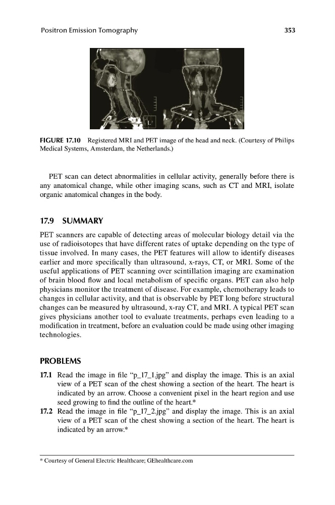
FIGURE 17.10 Registered MRI and PET image of the head and neck. (Courtesy of Philips
Medical Systems, Amsterdam, the Netherlands.)
PET scan can detect abnormalities in cellular activity, generally before there is
any anatomical change, while other imaging scans, such as CT and MRI, isolate
organic anatomical changes in the body.
17.9 SUMMARY
PET scanners are capable of detecting areas of molecular biology detail via the
use of radioisotopes that have different rates of uptake depending on the type of
tissue involved. In many cases, the PET features will allow to identify diseases
earlier and more specifically than ultrasound, x-rays, CT, or MRI. Some of the
useful applications of PET scanning over scintillation imaging are examination
of brain blood flow and local metabolism of specific organs. PET can also help
physicians monitor the treatment of disease. For example, chemotherapy leads to
changes in cellular activity, and that is observable by PET long before structural
changes can be measured by ultrasound, x-ray CT, and MRI. A typical PET scan
gives physicians another tool to evaluate treatments, perhaps even leading to a
modification in treatment, before an evaluation could be made using other imaging
technologies.
PROBLEMS
17.1 Read the image in file “p_17_1.jpg” and display the image. This is an axial
view of a PET scan of the chest showing a section of the heart. The heart is
indicated by an arrow. Choose a convenient pixel in the heart region and use
seed growing to find the outline of the heart.*
17.2 Read the image in file “p_17_2.jpg” and display the image. This is an axial
view of a PET scan of the chest showing a section of the heart. The heart is
indicated by an arrow.*
* Courtesy of General Electric Healthcare; GEhealthcare.com
L
A
Positron Emission Tomography
FIGURE 17.10 Registered MRI and PET image of the head and neck. (Courtesy of Philips
Medical Systems, Amsterdam, the Netherlands.)
PET scan can detect abnormalities in cellular activity, generally before there is
any anatomical change, while other imaging scans, such as CT and MRI, isolate
organic anatomical changes in the body.
17.9 SUMMARY
PET scanners are capable of detecting areas of molecular biology detail via the
use of radioisotopes that have different rates of uptake depending on the type of
tissue involved. In many cases, the PET features will allow to identify diseases
earlier and more specifically than ultrasound, x-rays, CT, or MRI. Some of the
useful applications of PET scanning over scintillation imaging are examination
of brain blood flow and local metabolism of specific organs. PET can also help
physicians monitor the treatment of disease. For example, chemotherapy leads to
changes in cellular activity, and that is observable by PET long before structural
changes can be measured by ultrasound, x-ray CT, and MRI. A typical PET scan
gives physicians another tool to evaluate treatments, perhaps even leading to a
modification in treatment, before an evaluation could be made using other imaging
technologies.
PROBLEMS
17.1 Read the image in file “p_17_1.jpg” and display the image. This is an axial
view of a PET scan of the chest showing a section of the heart. The heart is
indicated by an arrow. Choose a convenient pixel in the heart region and use
seed growing to find the outline of the heart.*
17.2 Read the image in file “p_17_2.jpg” and display the image. This is an axial
view of a PET scan of the chest showing a section of the heart. The heart is
indicated by an arrow.*
* Courtesy of General Electric Healthcare; GEhealthcare.com