

352
I
L
A
Biomedical Signal and Image Processing
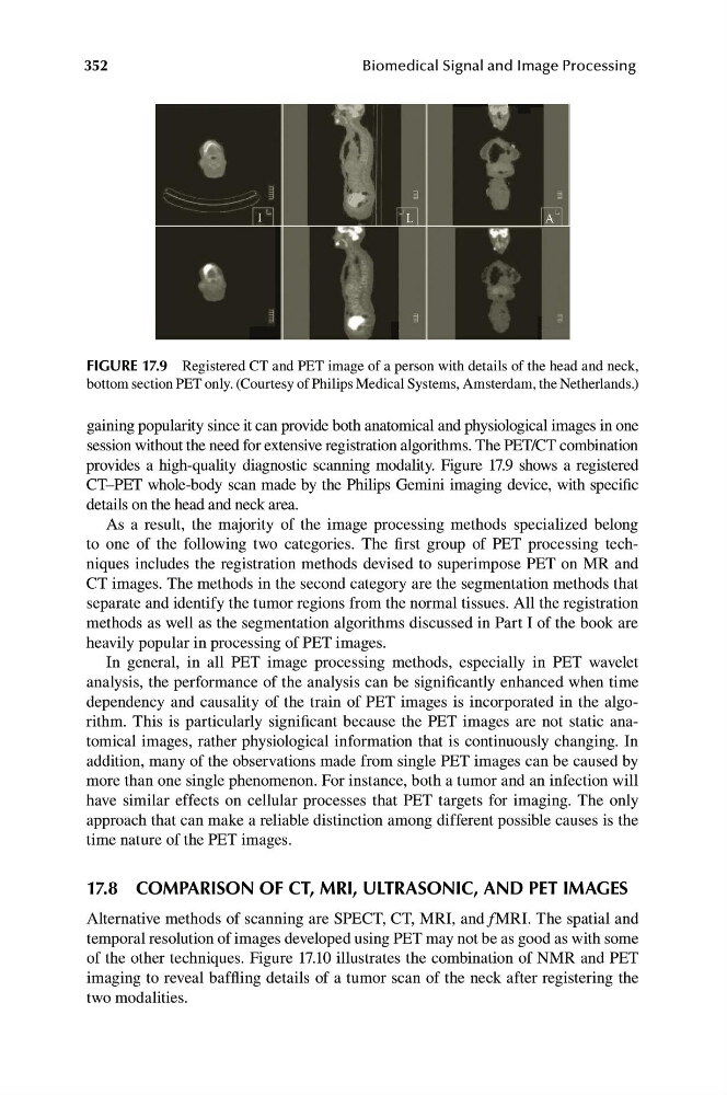
FIGURE 17.9 Registered CT and PET image of a person with details of the head and neck,
bottom section PET only. (Courtesy of Philips Medical Systems, Amsterdam, the Netherlands.)
gaining popularity since it can provide both anatomical and physiological images in one
session without the need for extensive registration algorithms. The PET/CT combination
provides a high-quality diagnostic scanning modality. Figure 17.9 shows a registered
CT–PET whole-body scan made by the Philips Gemini imaging device, with specific
details on the head and neck area.
As a result, the majority of the image processing methods specialized belong
to one of the following two categories. The first group of PET processing techniques includes the registration methods devised to superimpose PET on MR and
CT images. The methods in the second category are the segmentation methods that
separate and identify the tumor regions from the normal tissues. All the registration
methods as well as the segmentation algorithms discussed in Part I of the book are
heavily popular in processing of PET images.
In general, in all PET image processing methods, especially in PET wavelet
analysis, the performance of the analysis can be significantly enhanced when time
dependency and causality of the train of PET images is incorporated in the algorithm. This is particularly significant because the PET images are not static anatomical images, rather physiological information that is continuously changing. In
addition, many of the observations made from single PET images can be caused by
more than one single phenomenon. For instance, both a tumor and an infection will
have similar effects on cellular processes that PET targets for imaging. The only
approach that can make a reliable distinction among different possible causes is the
time nature of the PET images.
17.8 COMPARISON OF CT, MRI, ULTRASONIC, AND PET IMAGES
Alternative methods of scanning are SPECT, CT, MRI, and f MRI. The spatial and
temporal resolution of images developed using PET may not be as good as with some
of the other techniques. Figure 17.10 illustrates the combination of NMR and PET
imaging to reveal baffling details of a tumor scan of the neck after registering the
two modalities.
I
L
A
Biomedical Signal and Image Processing
FIGURE 17.9 Registered CT and PET image of a person with details of the head and neck,
bottom section PET only. (Courtesy of Philips Medical Systems, Amsterdam, the Netherlands.)
gaining popularity since it can provide both anatomical and physiological images in one
session without the need for extensive registration algorithms. The PET/CT combination
provides a high-quality diagnostic scanning modality. Figure 17.9 shows a registered
CT–PET whole-body scan made by the Philips Gemini imaging device, with specific
details on the head and neck area.
As a result, the majority of the image processing methods specialized belong
to one of the following two categories. The first group of PET processing techniques includes the registration methods devised to superimpose PET on MR and
CT images. The methods in the second category are the segmentation methods that
separate and identify the tumor regions from the normal tissues. All the registration
methods as well as the segmentation algorithms discussed in Part I of the book are
heavily popular in processing of PET images.
In general, in all PET image processing methods, especially in PET wavelet
analysis, the performance of the analysis can be significantly enhanced when time
dependency and causality of the train of PET images is incorporated in the algorithm. This is particularly significant because the PET images are not static anatomical images, rather physiological information that is continuously changing. In
addition, many of the observations made from single PET images can be caused by
more than one single phenomenon. For instance, both a tumor and an infection will
have similar effects on cellular processes that PET targets for imaging. The only
approach that can make a reliable distinction among different possible causes is the
time nature of the PET images.
17.8 COMPARISON OF CT, MRI, ULTRASONIC, AND PET IMAGES
Alternative methods of scanning are SPECT, CT, MRI, and f MRI. The spatial and
temporal resolution of images developed using PET may not be as good as with some
of the other techniques. Figure 17.10 illustrates the combination of NMR and PET
imaging to reveal baffling details of a tumor scan of the neck after registering the
two modalities.