

X-ray source
Rotation
only
Detectors
Biological
medium
275
X-Ray Imaging and Computed Tomography
Translation
X-ray source
Rotation
Biological
medium
Detectors
FIGURE 14.12 Diagram of second-generation tomography scanner utilizing the fan-beam
scanning principle, initially repeating the same linear scanning motion followed by rotation
as the first-generation tomograph. The fan beam however allows for a smaller dose due to
the collection of all transmitted radiation. (Courtesy of Dr. Bob Jones, Lancaster University,
Lancaster, United Kingdom. http://www.lancs.ac.uk/depts/physics/physics.htm)
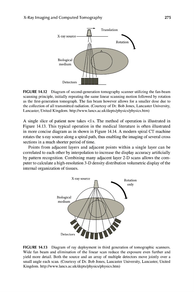
A single slice of patient now takes <1 s. The method of operation is illustrated in
Figure 14.13. This typical operation in the medical literature is often illustrated
in more concise diagram as in shown in Figure 14.14. A modern spiral CT machine
rotates the x-ray source along a spiral path, thus enabling the imaging of several cross
sections in a much shorter period of time.
Points from adjacent layers and adjacent points within a single layer can be
correlated to each other by interpolation to increase the display accuracy artificially
by pattern recognition. Combining many adjacent layer 2-D scans allows the computer to calculate a high-resolution 3-D density distribution volumetric display of the
internal organization of tissues.
FIGURE 14.13 Diagram of ray deployment in third generation of tomographic scanners.
Wide fan beam and elimination of the linear scan reduce the exposure even further and
yield more detail. Both the source and an array of multiple detectors move jointly over a
small angle each scan. (Courtesy of Dr. Bob Jones, Lancaster University, Lancaster, United
Kingdom. http://www.lancs.ac.uk/depts/physics/physics.htm)
Rotation
only
Detectors
Biological
medium
275
X-Ray Imaging and Computed Tomography
Translation
X-ray source
Rotation
Biological
medium
Detectors
FIGURE 14.12 Diagram of second-generation tomography scanner utilizing the fan-beam
scanning principle, initially repeating the same linear scanning motion followed by rotation
as the first-generation tomograph. The fan beam however allows for a smaller dose due to
the collection of all transmitted radiation. (Courtesy of Dr. Bob Jones, Lancaster University,
Lancaster, United Kingdom. http://www.lancs.ac.uk/depts/physics/physics.htm)
A single slice of patient now takes <1 s. The method of operation is illustrated in
Figure 14.13. This typical operation in the medical literature is often illustrated
in more concise diagram as in shown in Figure 14.14. A modern spiral CT machine
rotates the x-ray source along a spiral path, thus enabling the imaging of several cross
sections in a much shorter period of time.
Points from adjacent layers and adjacent points within a single layer can be
correlated to each other by interpolation to increase the display accuracy artificially
by pattern recognition. Combining many adjacent layer 2-D scans allows the computer to calculate a high-resolution 3-D density distribution volumetric display of the
internal organization of tissues.
FIGURE 14.13 Diagram of ray deployment in third generation of tomographic scanners.
Wide fan beam and elimination of the linear scan reduce the exposure even further and
yield more detail. Both the source and an array of multiple detectors move jointly over a
small angle each scan. (Courtesy of Dr. Bob Jones, Lancaster University, Lancaster, United
Kingdom. http://www.lancs.ac.uk/depts/physics/physics.htm)