

268
Biomedical Signal and Image Processing
Optical density (D) (arbitrary units)
3
D 2
2
D 1
1
Linear region
X 1
X 2 Log 10 X
Exposure (J/cm 2 )
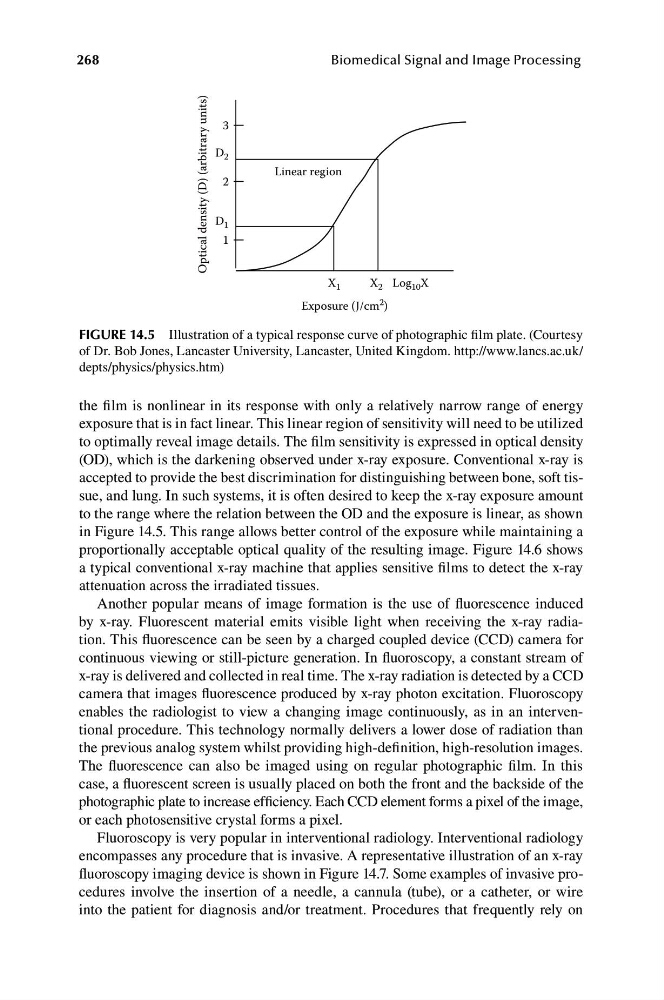
FIGURE 14.5 Illustration of a typical response curve of photographic film plate. (Courtesy
of Dr. Bob Jones, Lancaster University, Lancaster, United Kingdom. http://www.lancs.ac.uk/
depts/physics/physics.htm)
the film is nonlinear in its response with only a relatively narrow range of energy
exposure that is in fact linear. This linear region of sensitivity will need to be utilized
to optimally reveal image details. The film sensitivity is expressed in optical density
(OD), which is the darkening observed under x-ray exposure. Conventional x-ray is
accepted to provide the best discrimination for distinguishing between bone, soft tissue, and lung. In such systems, it is often desired to keep the x-ray exposure amount
to the range where the relation between the OD and the exposure is linear, as shown
in Figure 14.5. This range allows better control of the exposure while maintaining a
proportionally acceptable optical quality of the resulting image. Figure 14.6 shows
a typical conventional x-ray machine that applies sensitive films to detect the x-ray
attenuation across the irradiated tissues.
Another popular means of image formation is the use of fluorescence induced
by x-ray. Fluorescent material emits visible light when receiving the x-ray radiation. This fluorescence can be seen by a charged coupled device (CCD) camera for
continuous viewing or still-picture generation. In fluoroscopy, a constant stream of
x-ray is delivered and collected in real time. The x-ray radiation is detected by a CCD
camera that images fluorescence produced by x-ray photon excitation. Fluoroscopy
enables the radiologist to view a changing image continuously, as in an interventional procedure. This technology normally delivers a lower dose of radiation than
the previous analog system whilst providing high-definition, high-resolution images.
The fluorescence can also be imaged using on regular photographic film. In this
case, a fluorescent screen is usually placed on both the front and the backside of the
photographic plate to increase efficiency. Each CCD element forms a pixel of the image,
or each photosensitive crystal forms a pixel.
Fluoroscopy is very popular in interventional radiology. Interventional radiology
encompasses any procedure that is invasive. A representative illustration of an x-ray
fluoroscopy imaging device is shown in Figure 14.7. Some examples of invasive procedures involve the insertion of a needle, a cannula (tube), or a catheter, or wire
into the patient for diagnosis and/or treatment. Procedures that frequently rely on
Biomedical Signal and Image Processing
Optical density (D) (arbitrary units)
3
D 2
2
D 1
1
Linear region
X 1
X 2 Log 10 X
Exposure (J/cm 2 )
FIGURE 14.5 Illustration of a typical response curve of photographic film plate. (Courtesy
of Dr. Bob Jones, Lancaster University, Lancaster, United Kingdom. http://www.lancs.ac.uk/
depts/physics/physics.htm)
the film is nonlinear in its response with only a relatively narrow range of energy
exposure that is in fact linear. This linear region of sensitivity will need to be utilized
to optimally reveal image details. The film sensitivity is expressed in optical density
(OD), which is the darkening observed under x-ray exposure. Conventional x-ray is
accepted to provide the best discrimination for distinguishing between bone, soft tissue, and lung. In such systems, it is often desired to keep the x-ray exposure amount
to the range where the relation between the OD and the exposure is linear, as shown
in Figure 14.5. This range allows better control of the exposure while maintaining a
proportionally acceptable optical quality of the resulting image. Figure 14.6 shows
a typical conventional x-ray machine that applies sensitive films to detect the x-ray
attenuation across the irradiated tissues.
Another popular means of image formation is the use of fluorescence induced
by x-ray. Fluorescent material emits visible light when receiving the x-ray radiation. This fluorescence can be seen by a charged coupled device (CCD) camera for
continuous viewing or still-picture generation. In fluoroscopy, a constant stream of
x-ray is delivered and collected in real time. The x-ray radiation is detected by a CCD
camera that images fluorescence produced by x-ray photon excitation. Fluoroscopy
enables the radiologist to view a changing image continuously, as in an interventional procedure. This technology normally delivers a lower dose of radiation than
the previous analog system whilst providing high-definition, high-resolution images.
The fluorescence can also be imaged using on regular photographic film. In this
case, a fluorescent screen is usually placed on both the front and the backside of the
photographic plate to increase efficiency. Each CCD element forms a pixel of the image,
or each photosensitive crystal forms a pixel.
Fluoroscopy is very popular in interventional radiology. Interventional radiology
encompasses any procedure that is invasive. A representative illustration of an x-ray
fluoroscopy imaging device is shown in Figure 14.7. Some examples of invasive procedures involve the insertion of a needle, a cannula (tube), or a catheter, or wire
into the patient for diagnosis and/or treatment. Procedures that frequently rely on