

Bone
Muscle
Fat
100
10
1
0. 1
μ
ρ
—
Mass attenuation coeffi cient
10 keV
100 keV
1 MeV
10 MeV
Incident photon energy (J)
Greatest contrast
Most useful energy
between tissues for radionuclide imaging
for x-ray
267
X-Ray Imaging and Computed Tomography
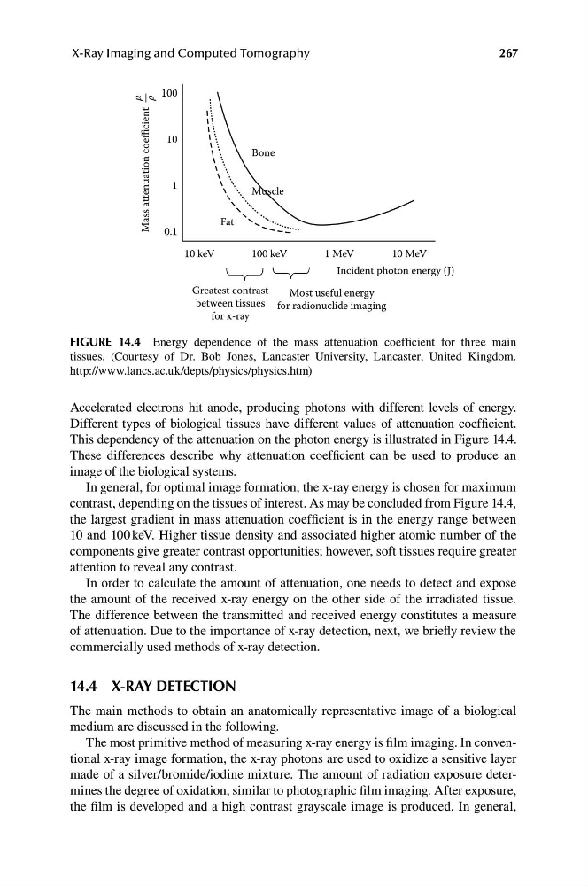
FIGURE 14.4 Energy dependence of the mass attenuation coefficient for three main
tissues. (Courtesy of Dr. Bob Jones, Lancaster University, Lancaster, United Kingdom.
http://www.lancs.ac.uk/depts/physics/physics.htm)
Accelerated electrons hit anode, producing photons with different levels of energy.
Different types of biological tissues have different values of attenuation coefficient.
This dependency of the attenuation on the photon energy is illustrated in Figure 14.4.
These differences describe why attenuation coefficient can be used to produce an
image of the biological systems.
In general, for optimal image formation, the x-ray energy is chosen for maximum
contrast, depending on the tissues of interest. As may be concluded from Figure 14.4,
the largest gradient in mass attenuation coefficient is in the energy range between
10 and 100 keV. Higher tissue density and associated higher atomic number of the
components give greater contrast opportunities; however, soft tissues require greater
attention to reveal any contrast.
In order to calculate the amount of attenuation, one needs to detect and expose
the amount of the received x-ray energy on the other side of the irradiated tissue.
The difference between the transmitted and received energy constitutes a measure
of attenuation. Due to the importance of x-ray detection, next, we briefly review the
commercially used methods of x-ray detection.
14.4 X-RAY DETECTION
The main methods to obtain an anatomically representative image of a biological
medium are discussed in the following.
The most primitive method of measuring x-ray energy is film imaging. In conventional x-ray image formation, the x-ray photons are used to oxidize a sensitive layer
made of a silver/bromide/iodine mixture. The amount of radiation exposure determines the degree of oxidation, similar to photographic film imaging. After exposure,
the film is developed and a high contrast grayscale image is produced. In general,
Muscle
Fat
100
10
1
0. 1
μ
ρ
—
Mass attenuation coeffi cient
10 keV
100 keV
1 MeV
10 MeV
Incident photon energy (J)
Greatest contrast
Most useful energy
between tissues for radionuclide imaging
for x-ray
267
X-Ray Imaging and Computed Tomography
FIGURE 14.4 Energy dependence of the mass attenuation coefficient for three main
tissues. (Courtesy of Dr. Bob Jones, Lancaster University, Lancaster, United Kingdom.
http://www.lancs.ac.uk/depts/physics/physics.htm)
Accelerated electrons hit anode, producing photons with different levels of energy.
Different types of biological tissues have different values of attenuation coefficient.
This dependency of the attenuation on the photon energy is illustrated in Figure 14.4.
These differences describe why attenuation coefficient can be used to produce an
image of the biological systems.
In general, for optimal image formation, the x-ray energy is chosen for maximum
contrast, depending on the tissues of interest. As may be concluded from Figure 14.4,
the largest gradient in mass attenuation coefficient is in the energy range between
10 and 100 keV. Higher tissue density and associated higher atomic number of the
components give greater contrast opportunities; however, soft tissues require greater
attention to reveal any contrast.
In order to calculate the amount of attenuation, one needs to detect and expose
the amount of the received x-ray energy on the other side of the irradiated tissue.
The difference between the transmitted and received energy constitutes a measure
of attenuation. Due to the importance of x-ray detection, next, we briefly review the
commercially used methods of x-ray detection.
14.4 X-RAY DETECTION
The main methods to obtain an anatomically representative image of a biological
medium are discussed in the following.
The most primitive method of measuring x-ray energy is film imaging. In conventional x-ray image formation, the x-ray photons are used to oxidize a sensitive layer
made of a silver/bromide/iodine mixture. The amount of radiation exposure determines the degree of oxidation, similar to photographic film imaging. After exposure,
the film is developed and a high contrast grayscale image is produced. In general,