

265
X-Ray Imaging and Computed Tomography
Primary photons
Source
Patient
Grid
Film
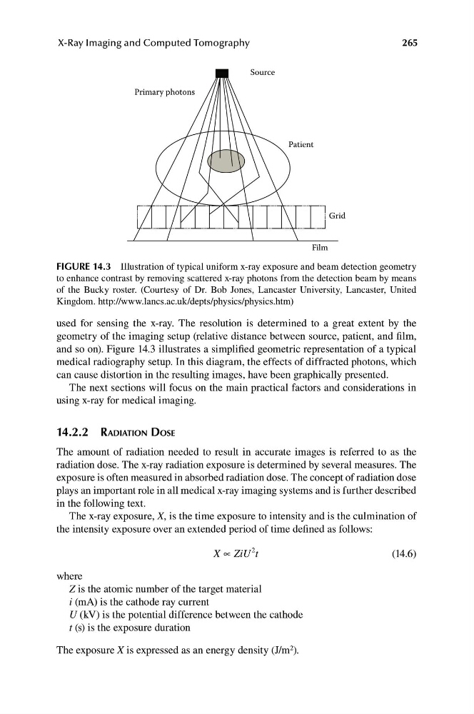
FIGURE 14.3 Illustration of typical uniform x-ray exposure and beam detection geometry
to enhance contrast by removing scattered x-ray photons from the detection beam by means
of the Bucky roster. (Courtesy of Dr. Bob Jones, Lancaster University, Lancaster, United
Kingdom. http://www.lancs.ac.uk/depts/physics/physics.htm)
used for sensing the x-ray. The resolution is determined to a great extent by the
geometry of the imaging setup (relative distance between source, patient, and film,
and so on). Figure 14.3 illustrates a simplified geometric representation of a typical
medical radiography setup. In this diagram, the effects of diffracted photons, which
can cause distortion in the resulting images, have been graphically presented.
The next sections will focus on the main practical factors and considerations in
using x-ray for medical imaging.
14.2.2 RADIATION DOSE
The amount of radiation needed to result in accurate images is referred to as the
radiation dose. The x-ray radiation exposure is determined by several measures. The
exposure is often measured in absorbed radiation dose. The concept of radiation dose
plays an important role in all medical x-ray imaging systems and is further described
in the following text.
The x-ray exposure, X, is the time exposure to intensity and is the culmination of
the intensity exposure over an extended period of time defined as follows:
X ∝ ZiU t
2
(14.6)
where
Z is the atomic number of the target material
i (mA) is the cathode ray current
U (kV) is the potential difference between the cathode
t (s) is the exposure duration
The exposure X is expressed as an energy density (J/m 2 ).
X-Ray Imaging and Computed Tomography
Primary photons
Source
Patient
Grid
Film
FIGURE 14.3 Illustration of typical uniform x-ray exposure and beam detection geometry
to enhance contrast by removing scattered x-ray photons from the detection beam by means
of the Bucky roster. (Courtesy of Dr. Bob Jones, Lancaster University, Lancaster, United
Kingdom. http://www.lancs.ac.uk/depts/physics/physics.htm)
used for sensing the x-ray. The resolution is determined to a great extent by the
geometry of the imaging setup (relative distance between source, patient, and film,
and so on). Figure 14.3 illustrates a simplified geometric representation of a typical
medical radiography setup. In this diagram, the effects of diffracted photons, which
can cause distortion in the resulting images, have been graphically presented.
The next sections will focus on the main practical factors and considerations in
using x-ray for medical imaging.
14.2.2 RADIATION DOSE
The amount of radiation needed to result in accurate images is referred to as the
radiation dose. The x-ray radiation exposure is determined by several measures. The
exposure is often measured in absorbed radiation dose. The concept of radiation dose
plays an important role in all medical x-ray imaging systems and is further described
in the following text.
The x-ray exposure, X, is the time exposure to intensity and is the culmination of
the intensity exposure over an extended period of time defined as follows:
X ∝ ZiU t
2
(14.6)
where
Z is the atomic number of the target material
i (mA) is the cathode ray current
U (kV) is the potential difference between the cathode
t (s) is the exposure duration
The exposure X is expressed as an energy density (J/m 2 ).