

Characteristic radiation
Acceleration voltage
200 kV
Bremsstrahlung
50 kV
anode voltage
Photon energy keV
Radiation exposure (J/m
2
)
264
Biomedical Signal and Image Processing
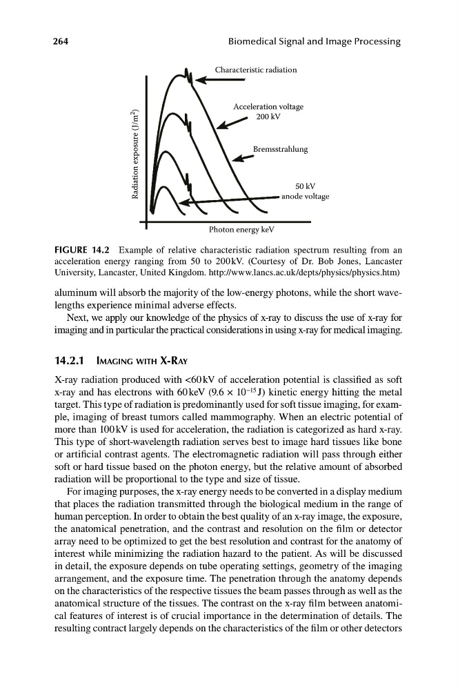
FIGURE 14.2 Example of relative characteristic radiation spectrum resulting from an
acceleration energy ranging from 50 to 200 kV. (Courtesy of Dr. Bob Jones, Lancaster
University, Lancaster, United Kingdom. http://www.lancs.ac.uk/depts/physics/physics.htm)
aluminum will absorb the majority of the low-energy photons, while the short wavelengths experience minimal adverse effects.
Next, we apply our knowledge of the physics of x-ray to discuss the use of x-ray for
imaging and in particular the practical considerations in using x-ray for medical imaging.
14.2.1 IMAGING WITH X-RAY
X-ray radiation produced with <60 kV of acceleration potential is classified as soft
x-ray and has electrons with 60 keV (9.6 × 10 −15 J) kinetic energy hitting the metal
target. This type of radiation is predominantly used for soft tissue imaging, for example, imaging of breast tumors called mammography. When an electric potential of
more than 100 kV is used for acceleration, the radiation is categorized as hard x-ray.
This type of short-wavelength radiation serves best to image hard tissues like bone
or artificial contrast agents. The electromagnetic radiation will pass through either
soft or hard tissue based on the photon energy, but the relative amount of absorbed
radiation will be proportional to the type and size of tissue.
For imaging purposes, the x-ray energy needs to be converted in a display medium
that places the radiation transmitted through the biological medium in the range of
human perception. In order to obtain the best quality of an x-ray image, the exposure,
the anatomical penetration, and the contrast and resolution on the film or detector
array need to be optimized to get the best resolution and contrast for the anatomy of
interest while minimizing the radiation hazard to the patient. As will be discussed
in detail, the exposure depends on tube operating settings, geometry of the imaging
arrangement, and the exposure time. The penetration through the anatomy depends
on the characteristics of the respective tissues the beam passes through as well as the
anatomical structure of the tissues. The contrast on the x-ray film between anatomical features of interest is of crucial importance in the determination of details. The
resulting contract largely depends on the characteristics of the film or other detectors
Acceleration voltage
200 kV
Bremsstrahlung
50 kV
anode voltage
Photon energy keV
Radiation exposure (J/m
2
)
264
Biomedical Signal and Image Processing
FIGURE 14.2 Example of relative characteristic radiation spectrum resulting from an
acceleration energy ranging from 50 to 200 kV. (Courtesy of Dr. Bob Jones, Lancaster
University, Lancaster, United Kingdom. http://www.lancs.ac.uk/depts/physics/physics.htm)
aluminum will absorb the majority of the low-energy photons, while the short wavelengths experience minimal adverse effects.
Next, we apply our knowledge of the physics of x-ray to discuss the use of x-ray for
imaging and in particular the practical considerations in using x-ray for medical imaging.
14.2.1 IMAGING WITH X-RAY
X-ray radiation produced with <60 kV of acceleration potential is classified as soft
x-ray and has electrons with 60 keV (9.6 × 10 −15 J) kinetic energy hitting the metal
target. This type of radiation is predominantly used for soft tissue imaging, for example, imaging of breast tumors called mammography. When an electric potential of
more than 100 kV is used for acceleration, the radiation is categorized as hard x-ray.
This type of short-wavelength radiation serves best to image hard tissues like bone
or artificial contrast agents. The electromagnetic radiation will pass through either
soft or hard tissue based on the photon energy, but the relative amount of absorbed
radiation will be proportional to the type and size of tissue.
For imaging purposes, the x-ray energy needs to be converted in a display medium
that places the radiation transmitted through the biological medium in the range of
human perception. In order to obtain the best quality of an x-ray image, the exposure,
the anatomical penetration, and the contrast and resolution on the film or detector
array need to be optimized to get the best resolution and contrast for the anatomy of
interest while minimizing the radiation hazard to the patient. As will be discussed
in detail, the exposure depends on tube operating settings, geometry of the imaging
arrangement, and the exposure time. The penetration through the anatomy depends
on the characteristics of the respective tissues the beam passes through as well as the
anatomical structure of the tissues. The contrast on the x-ray film between anatomical features of interest is of crucial importance in the determination of details. The
resulting contract largely depends on the characteristics of the film or other detectors