

250
Biomedical Signal and Image Processing
(e.g., estimating the internal structure of a crystal without breaking it), remote sensing
(e.g., using satellite image to estimate the shape and distribution of weather fronts), and
mining (e.g., discovering the existence of a particular mineral inside a mountain or deep
under the ground).
Returning to our discussion of CT for biomedical imaging, from the standpoint
of how the system is stimulated and how the measurements are made, CT systems
can be categorized into three most commonly used types: attenuation tomography,
reflection tomography, and refraction tomography. A brief and high-level description
of these three approaches that avoids any mathematical formulations is given in the
following.
13.1.1 ATTENUATION TOMOGRAPHY
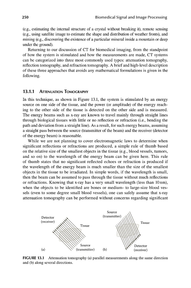
In this technique, as shown in Figure 13.1, the system is stimulated by an energy
source on one side of the tissue, and the power (or amplitude) of the energy reaching to the other side of the tissue is detected on the other side and is measured.
The energy beams such as x-ray are known to travel mainly through straight lines
through biological tissues with little or no reflection or refraction (i.e., bending the
path and deviation from a straight line). As a result, for such energy beams, assuming
a straight pass between the source (transmitter of the beam) and the receiver (detector
of the energy beam) is reasonable.
While we are not planning to cover electromagnetic laws to determine when
significant reflections or refractions are produced, a simple rule of thumb based
on the relative size of the smallest objects in the tissue (e.g., blood vessels, tumors,
and so on) to the wavelength of the energy beam can be given here. This rule
of thumb states that no significant reflected echoes or refraction is produced if
the wavelength of the energy beam is much smaller than the size of the smallest
objects in the tissue to be irradiated. In simple words, if the wavelength is small,
then the beam can be assumed to pass through the tissue without much reflections
or refractions. Knowing that x-ray has a very small wavelength (less than 10 nm),
when the objects to be identified are bones or medium- to large-size blood vessels (even to some degree small blood vessels), one can safely assume that x-ray
attenuation tomography can be performed without concerns regarding significant
Source
Detector
(transmitter)
Detector
Tissue
Source
(receiver)
Tissue
(a)
(transmitter)
(b)
(receiver)
FIGURE 13.1 Attenuation tomography (a) parallel measurements along the same direction
and (b) along several directions.
Biomedical Signal and Image Processing
(e.g., estimating the internal structure of a crystal without breaking it), remote sensing
(e.g., using satellite image to estimate the shape and distribution of weather fronts), and
mining (e.g., discovering the existence of a particular mineral inside a mountain or deep
under the ground).
Returning to our discussion of CT for biomedical imaging, from the standpoint
of how the system is stimulated and how the measurements are made, CT systems
can be categorized into three most commonly used types: attenuation tomography,
reflection tomography, and refraction tomography. A brief and high-level description
of these three approaches that avoids any mathematical formulations is given in the
following.
13.1.1 ATTENUATION TOMOGRAPHY
In this technique, as shown in Figure 13.1, the system is stimulated by an energy
source on one side of the tissue, and the power (or amplitude) of the energy reaching to the other side of the tissue is detected on the other side and is measured.
The energy beams such as x-ray are known to travel mainly through straight lines
through biological tissues with little or no reflection or refraction (i.e., bending the
path and deviation from a straight line). As a result, for such energy beams, assuming
a straight pass between the source (transmitter of the beam) and the receiver (detector
of the energy beam) is reasonable.
While we are not planning to cover electromagnetic laws to determine when
significant reflections or refractions are produced, a simple rule of thumb based
on the relative size of the smallest objects in the tissue (e.g., blood vessels, tumors,
and so on) to the wavelength of the energy beam can be given here. This rule
of thumb states that no significant reflected echoes or refraction is produced if
the wavelength of the energy beam is much smaller than the size of the smallest
objects in the tissue to be irradiated. In simple words, if the wavelength is small,
then the beam can be assumed to pass through the tissue without much reflections
or refractions. Knowing that x-ray has a very small wavelength (less than 10 nm),
when the objects to be identified are bones or medium- to large-size blood vessels (even to some degree small blood vessels), one can safely assume that x-ray
attenuation tomography can be performed without concerns regarding significant
Source
Detector
(transmitter)
Detector
Tissue
Source
(receiver)
Tissue
(a)
(transmitter)
(b)
(receiver)
FIGURE 13.1 Attenuation tomography (a) parallel measurements along the same direction
and (b) along several directions.