

242
Biomedical Signal and Image Processing
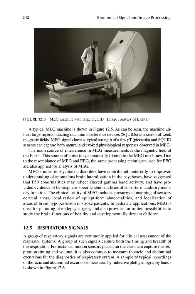
FIGURE 12.5 MEG machine with large SQUID. (Image courtesy of Elekta.)
A typical MEG machine is shown in Figure 12.5. As can be seen, the machine utilizes large superconducting quantum interference devices (SQUIDs) as a sensor of weak
magnetic fields. MEG signals have a typical strength of a few pT (picotesla) and SQUID
sensors can capture both natural and evoked physiological responses observed in MEG.
The main source of interference in MEG measurements is the magnetic field of
the Earth. This source of noise is systematically filtered in the MEG machines. Due
to the resemblance of MEG and EEG, the same processing techniques used for EEG
are also applied for analysis of MEG.
MEG studies in psychiatric disorders have contributed materially to improved
understanding of anomalous brain lateralization in the psychoses, have suggested
that P50 abnormalities may reflect altered gamma band activity, and have provided evidence of hemisphere-specific abnormalities of short-term auditory memory function. The clinical utility of MEG includes presurgical mapping of sensory
cortical areas, localization of epileptiform abnormalities, and localization of
areas of brain hypoperfusion in stroke patients. In pediatric applications, MEG is
used for planning of epilepsy surgery and also provides unlimited possibilities to
study the brain functions of healthy and developmentally deviant children.
12.5 RESPIRATORY SIGNALS
A group of respiratory signals are commonly applied for clinical assessment of the
respirator systems. A group of such signals capture both the timing and breadth of
the respiration. For instance, motion sensors placed on the chest can capture the respiration timing and volume. It is also common to measure thoracic and abdominal
excursions for the diagnostics of respiratory system. A sample of typical recordings
of thoracic and abdominal excursions measured by inductive plethysmography bands
is shown in Figure 12.6.
Biomedical Signal and Image Processing
FIGURE 12.5 MEG machine with large SQUID. (Image courtesy of Elekta.)
A typical MEG machine is shown in Figure 12.5. As can be seen, the machine utilizes large superconducting quantum interference devices (SQUIDs) as a sensor of weak
magnetic fields. MEG signals have a typical strength of a few pT (picotesla) and SQUID
sensors can capture both natural and evoked physiological responses observed in MEG.
The main source of interference in MEG measurements is the magnetic field of
the Earth. This source of noise is systematically filtered in the MEG machines. Due
to the resemblance of MEG and EEG, the same processing techniques used for EEG
are also applied for analysis of MEG.
MEG studies in psychiatric disorders have contributed materially to improved
understanding of anomalous brain lateralization in the psychoses, have suggested
that P50 abnormalities may reflect altered gamma band activity, and have provided evidence of hemisphere-specific abnormalities of short-term auditory memory function. The clinical utility of MEG includes presurgical mapping of sensory
cortical areas, localization of epileptiform abnormalities, and localization of
areas of brain hypoperfusion in stroke patients. In pediatric applications, MEG is
used for planning of epilepsy surgery and also provides unlimited possibilities to
study the brain functions of healthy and developmentally deviant children.
12.5 RESPIRATORY SIGNALS
A group of respiratory signals are commonly applied for clinical assessment of the
respirator systems. A group of such signals capture both the timing and breadth of
the respiration. For instance, motion sensors placed on the chest can capture the respiration timing and volume. It is also common to measure thoracic and abdominal
excursions for the diagnostics of respiratory system. A sample of typical recordings
of thoracic and abdominal excursions measured by inductive plethysmography bands
is shown in Figure 12.6.