

224
Biomedical Signal and Image Processing
In such EMG recordings, electrodes incorporated in very fine needles can be inserted
in the muscle itself. These electrodes record the electric potential generated by the
depolarization of the muscle cells directly surrounding the needle electrode. More
specifically, in such neuromuscular measurements, the electric activity of a single
motor unit is directly measured. When a needle has more than one electrode, bipolar measurements can be made to derive potential gradients within the muscle.
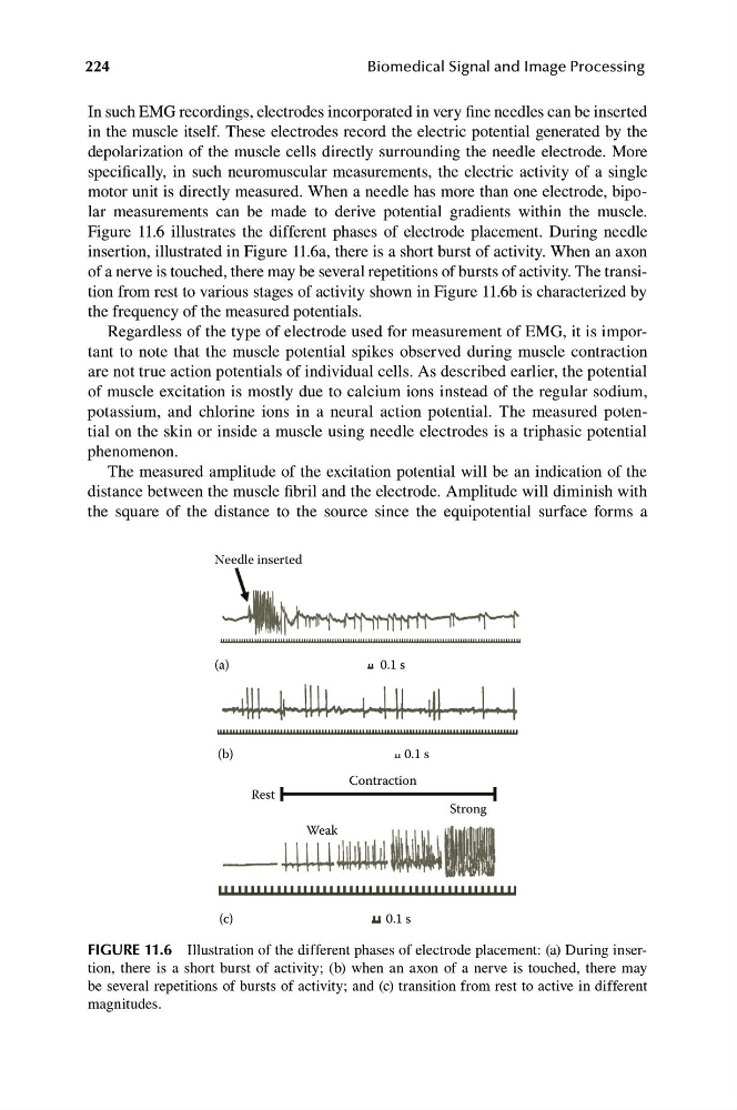
Figure 11.6 illustrates the different phases of electrode placement. During needle
insertion, illustrated in Figure 11.6a, there is a short burst of activity. When an axon
of a nerve is touched, there may be several repetitions of bursts of activity. The transition from rest to various stages of activity shown in Figure 11.6b is characterized by
the frequency of the measured potentials.
Regardless of the type of electrode used for measurement of EMG, it is important to note that the muscle potential spikes observed during muscle contraction
are not true action potentials of individual cells. As described earlier, the potential
of muscle excitation is mostly due to calcium ions instead of the regular sodium,
potassium, and chlorine ions in a neural action potential. The measured potential on the skin or inside a muscle using needle electrodes is a triphasic potential
phenomenon.
The measured amplitude of the excitation potential will be an indication of the
distance between the muscle fibril and the electrode. Amplitude will diminish with
the square of the distance to the source since the equipotential surface forms a
Needle inserted
0.1 s
(a)
0.1 s
(b)
Contraction
0.1 s
(c)
Weak
Strong
Rest
FIGURE 11.6 Illustration of the different phases of electrode placement: (a) During insertion, there is a short burst of activity; (b) when an axon of a nerve is touched, there may
be several repetitions of bursts of activity; and (c) transition from rest to active in different
magnitudes.
Biomedical Signal and Image Processing
In such EMG recordings, electrodes incorporated in very fine needles can be inserted
in the muscle itself. These electrodes record the electric potential generated by the
depolarization of the muscle cells directly surrounding the needle electrode. More
specifically, in such neuromuscular measurements, the electric activity of a single
motor unit is directly measured. When a needle has more than one electrode, bipolar measurements can be made to derive potential gradients within the muscle.
Figure 11.6 illustrates the different phases of electrode placement. During needle
insertion, illustrated in Figure 11.6a, there is a short burst of activity. When an axon
of a nerve is touched, there may be several repetitions of bursts of activity. The transition from rest to various stages of activity shown in Figure 11.6b is characterized by
the frequency of the measured potentials.
Regardless of the type of electrode used for measurement of EMG, it is important to note that the muscle potential spikes observed during muscle contraction
are not true action potentials of individual cells. As described earlier, the potential
of muscle excitation is mostly due to calcium ions instead of the regular sodium,
potassium, and chlorine ions in a neural action potential. The measured potential on the skin or inside a muscle using needle electrodes is a triphasic potential
phenomenon.
The measured amplitude of the excitation potential will be an indication of the
distance between the muscle fibril and the electrode. Amplitude will diminish with
the square of the distance to the source since the equipotential surface forms a
Needle inserted
0.1 s
(a)
0.1 s
(b)
Contraction
0.1 s
(c)
Weak
Strong
Rest
FIGURE 11.6 Illustration of the different phases of electrode placement: (a) During insertion, there is a short burst of activity; (b) when an axon of a nerve is touched, there may
be several repetitions of bursts of activity; and (c) transition from rest to active in different
magnitudes.