

303
606
908 1210 1513
Sample at 60,000 Hz
2.76
1.28
–0.20
–1.68
–3.16
1
Amplitude (arbitrary units)
1815 2117 2419 2722 3024
Electromyogram
223
11.3 EMG: SIGNAL OF MUSCLES
There are two general methods of recording the electric activities of muscle tissue.
One method applies electrodes on the skin and records a surface EMG. The second
method actually inserts needles with electrodes into the muscle itself.
In most applications, what we measure as EMG is the spatially weighted sum of
the electric activities of a number of motor units that are collected on the surface
of the skin. In other words, a typical EMG signal is the resultant of the electric
activities of many motor units that are weighted according to the amount of the
fat and skin between each motor unit and the electrode as well as the distance of
each motor unit from the location of the electrode. Electrodes placed on the skin
surface can be used to monitor the coordination of entire muscle groups; however,
not much will be known about the individual muscle cells from this. In general,
this technique is used to identify which muscle groups are involved in a particular
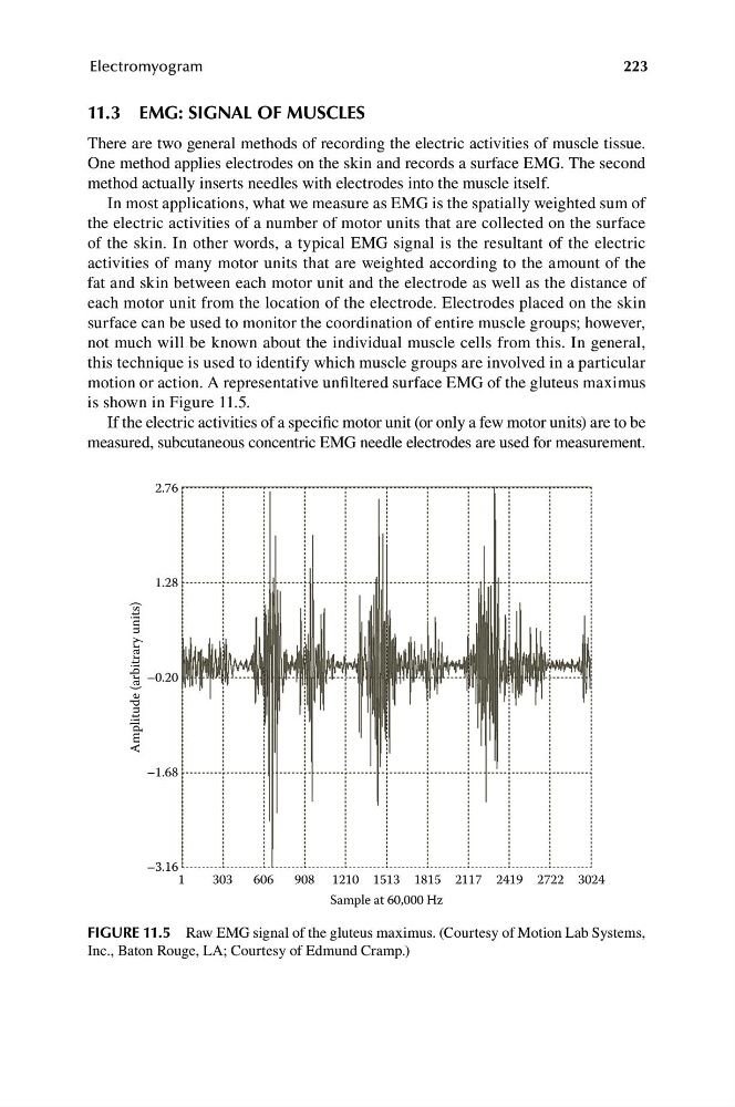
motion or action. A representative unfiltered surface EMG of the gluteus maximus
is shown in Figure 11.5.
If the electric activities of a specific motor unit (or only a few motor units) are to be
measured, subcutaneous concentric EMG needle electrodes are used for measurement.
FIGURE 11.5 Raw EMG signal of the gluteus maximus. (Courtesy of Motion Lab Systems,
Inc., Baton Rouge, LA; Courtesy of Edmund Cramp.)
606
908 1210 1513
Sample at 60,000 Hz
2.76
1.28
–0.20
–1.68
–3.16
1
Amplitude (arbitrary units)
1815 2117 2419 2722 3024
Electromyogram
223
11.3 EMG: SIGNAL OF MUSCLES
There are two general methods of recording the electric activities of muscle tissue.
One method applies electrodes on the skin and records a surface EMG. The second
method actually inserts needles with electrodes into the muscle itself.
In most applications, what we measure as EMG is the spatially weighted sum of
the electric activities of a number of motor units that are collected on the surface
of the skin. In other words, a typical EMG signal is the resultant of the electric
activities of many motor units that are weighted according to the amount of the
fat and skin between each motor unit and the electrode as well as the distance of
each motor unit from the location of the electrode. Electrodes placed on the skin
surface can be used to monitor the coordination of entire muscle groups; however,
not much will be known about the individual muscle cells from this. In general,
this technique is used to identify which muscle groups are involved in a particular
motion or action. A representative unfiltered surface EMG of the gluteus maximus
is shown in Figure 11.5.
If the electric activities of a specific motor unit (or only a few motor units) are to be
measured, subcutaneous concentric EMG needle electrodes are used for measurement.
FIGURE 11.5 Raw EMG signal of the gluteus maximus. (Courtesy of Motion Lab Systems,
Inc., Baton Rouge, LA; Courtesy of Edmund Cramp.)