

220
Myosin filament
Ca 2+
Ca
2+
Ca
2+
Ca
2+
Ca
2+
Ca
2+
Ca 2+
A-band
Actin filament
Z-disk
M-disk
Biomedical Signal and Image Processing
there are rather larger muscles in charge of relatively coarse motions. In these large
muscles, which include the large muscle of the lower limb such as gluteus maximus,
each motor unit engages approximately 2000 muscle fibers.
In order to see how contraction is performed by a muscle, we need to focus on the
internal structure of a typical muscle and describe the interaction among the molecules involved in the process of contraction.
11.2.2 MUSCLE CONTRACTION
A muscle is made up of several muscle cells, and every muscle cell has a larger number of muscle fibers. Each muscle fiber is made of strands of tissues, called fibrils
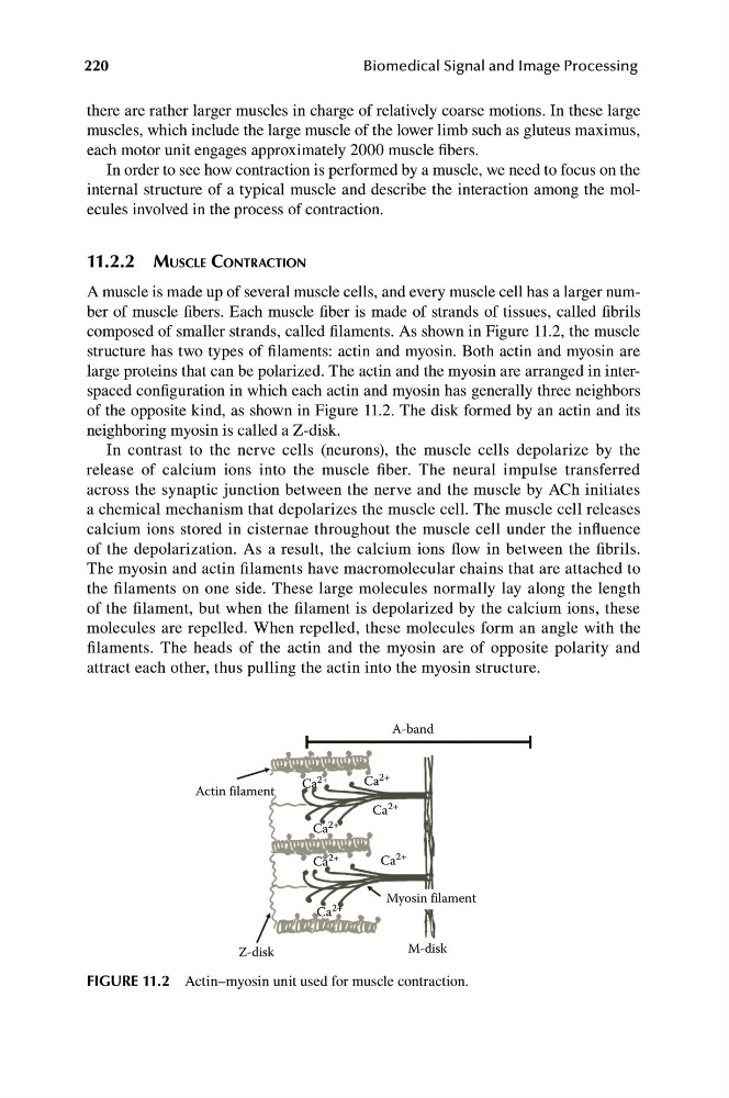
composed of smaller strands, called filaments. As shown in Figure 11.2, the muscle
structure has two types of filaments: actin and myosin. Both actin and myosin are
large proteins that can be polarized. The actin and the myosin are arranged in interspaced configuration in which each actin and myosin has generally three neighbors
of the opposite kind, as shown in Figure 11.2. The disk formed by an actin and its
neighboring myosin is called a Z-disk.
In contrast to the nerve cells (neurons), the muscle cells depolarize by the
release of calcium ions into the muscle fiber. The neural impulse transferred
across the synaptic junction between the nerve and the muscle by ACh initiates
a chemical mechanism that depolarizes the muscle cell. The muscle cell releases
calcium ions stored in cisternae throughout the muscle cell under the influence
of the depolarization. As a result, the calcium ions flow in between the fibrils.
The myosin and actin filaments have macromolecular chains that are attached to
the filaments on one side. These large molecules normally lay along the length
of the filament, but when the filament is depolarized by the calcium ions, these
molecules are repelled. When repelled, these molecules form an angle with the
filaments. The heads of the actin and the myosin are of opposite polarity and
attract each other, thus pulling the actin into the myosin structure.
FIGURE 11.2 Actin–myosin unit used for muscle contraction.
Myosin filament
Ca 2+
Ca
2+
Ca
2+
Ca
2+
Ca
2+
Ca
2+
Ca 2+
A-band
Actin filament
Z-disk
M-disk
Biomedical Signal and Image Processing
there are rather larger muscles in charge of relatively coarse motions. In these large
muscles, which include the large muscle of the lower limb such as gluteus maximus,
each motor unit engages approximately 2000 muscle fibers.
In order to see how contraction is performed by a muscle, we need to focus on the
internal structure of a typical muscle and describe the interaction among the molecules involved in the process of contraction.
11.2.2 MUSCLE CONTRACTION
A muscle is made up of several muscle cells, and every muscle cell has a larger number of muscle fibers. Each muscle fiber is made of strands of tissues, called fibrils
composed of smaller strands, called filaments. As shown in Figure 11.2, the muscle
structure has two types of filaments: actin and myosin. Both actin and myosin are
large proteins that can be polarized. The actin and the myosin are arranged in interspaced configuration in which each actin and myosin has generally three neighbors
of the opposite kind, as shown in Figure 11.2. The disk formed by an actin and its
neighboring myosin is called a Z-disk.
In contrast to the nerve cells (neurons), the muscle cells depolarize by the
release of calcium ions into the muscle fiber. The neural impulse transferred
across the synaptic junction between the nerve and the muscle by ACh initiates
a chemical mechanism that depolarizes the muscle cell. The muscle cell releases
calcium ions stored in cisternae throughout the muscle cell under the influence
of the depolarization. As a result, the calcium ions flow in between the fibrils.
The myosin and actin filaments have macromolecular chains that are attached to
the filaments on one side. These large molecules normally lay along the length
of the filament, but when the filament is depolarized by the calcium ions, these
molecules are repelled. When repelled, these molecules form an angle with the
filaments. The heads of the actin and the myosin are of opposite polarity and
attract each other, thus pulling the actin into the myosin structure.
FIGURE 11.2 Actin–myosin unit used for muscle contraction.