

208
Biomedical Signal and Image Processing
The focal location of the origin of the epileptic signal is usually determined by
finding the inverse solution to a so-called equivalent dipole source from the EEG
recordings. The equivalent dipole source feature was described in Chapter 8.
Combining the spatial distribution with the temporal information of the representative features in the EEG signal from many electrodes can usually resolve the source
location with relatively high degree of accuracy.
10.5.2 SLEEP DISORDERS
Due to the distinguished differences in frequency content of the awake and sleep
EEG, the EEG recordings are heavily used in diagnosing sleep disorders. As mentioned earlier in this chapter, the EEG of a person at rest is in the low-frequency
ranges, especially with the eyes closed. However, there are different stages in sleep
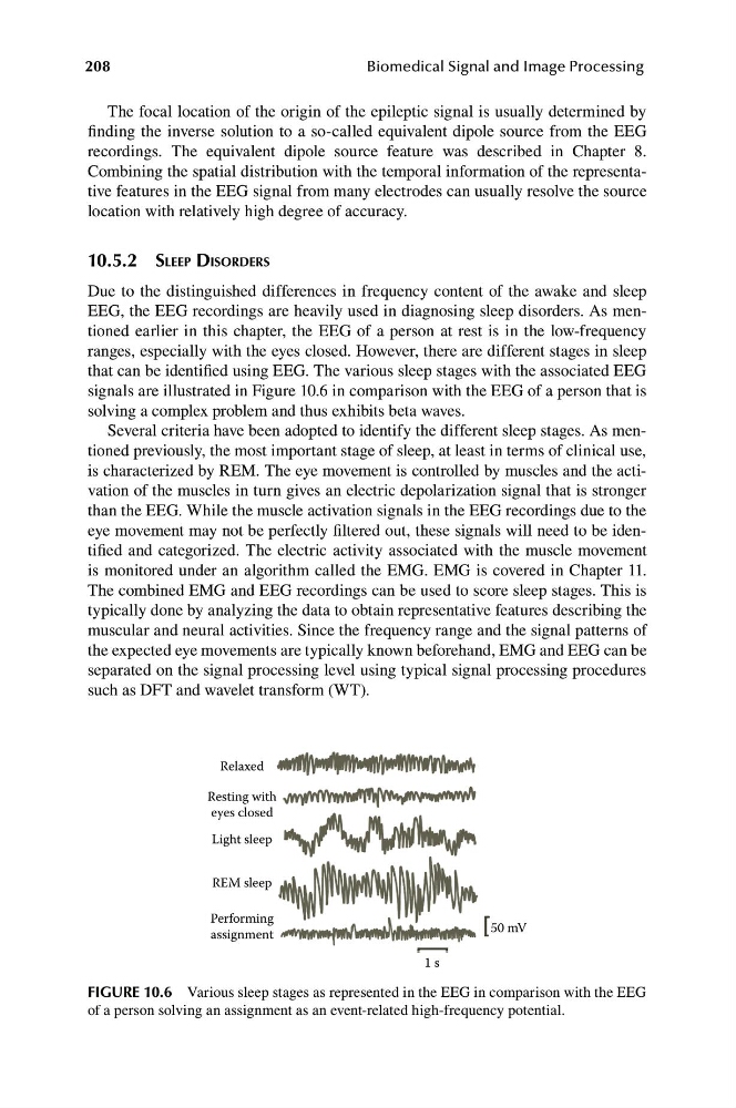
that can be identified using EEG. The various sleep stages with the associated EEG
signals are illustrated in Figure 10.6 in comparison with the EEG of a person that is
solving a complex problem and thus exhibits beta waves.
Several criteria have been adopted to identify the different sleep stages. As mentioned previously, the most important stage of sleep, at least in terms of clinical use,
is characterized by REM. The eye movement is controlled by muscles and the activation of the muscles in turn gives an electric depolarization signal that is stronger
than the EEG. While the muscle activation signals in the EEG recordings due to the
eye movement may not be perfectly filtered out, these signals will need to be identified and categorized. The electric activity associated with the muscle movement
is monitored under an algorithm called the EMG. EMG is covered in Chapter 11.
The combined EMG and EEG recordings can be used to score sleep stages. This is
typically done by analyzing the data to obtain representative features describing the
muscular and neural activities. Since the frequency range and the signal patterns of
the expected eye movements are typically known beforehand, EMG and EEG can be
separated on the signal processing level using typical signal processing procedures
such as DFT and wavelet transform (WT).
Relaxed
Resting with
eyes closed
Light sleep
REM sleep
Performing
50 mV
assignment
1 s
FIGURE 10.6 Various sleep stages as represented in the EEG in comparison with the EEG
of a person solving an assignment as an event-related high-frequency potential.
Biomedical Signal and Image Processing
The focal location of the origin of the epileptic signal is usually determined by
finding the inverse solution to a so-called equivalent dipole source from the EEG
recordings. The equivalent dipole source feature was described in Chapter 8.
Combining the spatial distribution with the temporal information of the representative features in the EEG signal from many electrodes can usually resolve the source
location with relatively high degree of accuracy.
10.5.2 SLEEP DISORDERS
Due to the distinguished differences in frequency content of the awake and sleep
EEG, the EEG recordings are heavily used in diagnosing sleep disorders. As mentioned earlier in this chapter, the EEG of a person at rest is in the low-frequency
ranges, especially with the eyes closed. However, there are different stages in sleep
that can be identified using EEG. The various sleep stages with the associated EEG
signals are illustrated in Figure 10.6 in comparison with the EEG of a person that is
solving a complex problem and thus exhibits beta waves.
Several criteria have been adopted to identify the different sleep stages. As mentioned previously, the most important stage of sleep, at least in terms of clinical use,
is characterized by REM. The eye movement is controlled by muscles and the activation of the muscles in turn gives an electric depolarization signal that is stronger
than the EEG. While the muscle activation signals in the EEG recordings due to the
eye movement may not be perfectly filtered out, these signals will need to be identified and categorized. The electric activity associated with the muscle movement
is monitored under an algorithm called the EMG. EMG is covered in Chapter 11.
The combined EMG and EEG recordings can be used to score sleep stages. This is
typically done by analyzing the data to obtain representative features describing the
muscular and neural activities. Since the frequency range and the signal patterns of
the expected eye movements are typically known beforehand, EMG and EEG can be
separated on the signal processing level using typical signal processing procedures
such as DFT and wavelet transform (WT).
Relaxed
Resting with
eyes closed
Light sleep
REM sleep
Performing
50 mV
assignment
1 s
FIGURE 10.6 Various sleep stages as represented in the EEG in comparison with the EEG
of a person solving an assignment as an event-related high-frequency potential.