

182
Biomedical Signal and Image Processing
9.4 CARDIOVASCULAR DISEASES AND ECG
These deviations from the normal functionality of the cardiovascular system are
associated with certain pathological conditions, which can be either genetic or due
to malfunctions such as infections, lack of oxygen, and obstruction of blood vessels
that supply blood to the heart itself. In this section, some of the main cardiovascular
diseases are briefly introduced and the changes in ECG and related diseases are
discussed. This brief discussion of cardiovascular diseases will allow us to devise
computational methods for the processing of ECG that can detect abnormalities in
their early stages.
9.4.1 ATRIAL FIBRILLATION
AF is one of the most common arrhythmias that occur as a result of rheumatic disease, infections (such as pericarditis), atherosclerotic disease, or hyperthyroidism.
These conditions caused by AF are not as life threatening as some of the ventricular
arrhythmias but provide an increased risk for stroke. Physical symptoms include
palpitations, dizziness, and shortness of breath. Some people having AF never notice
any sign of discomfort.
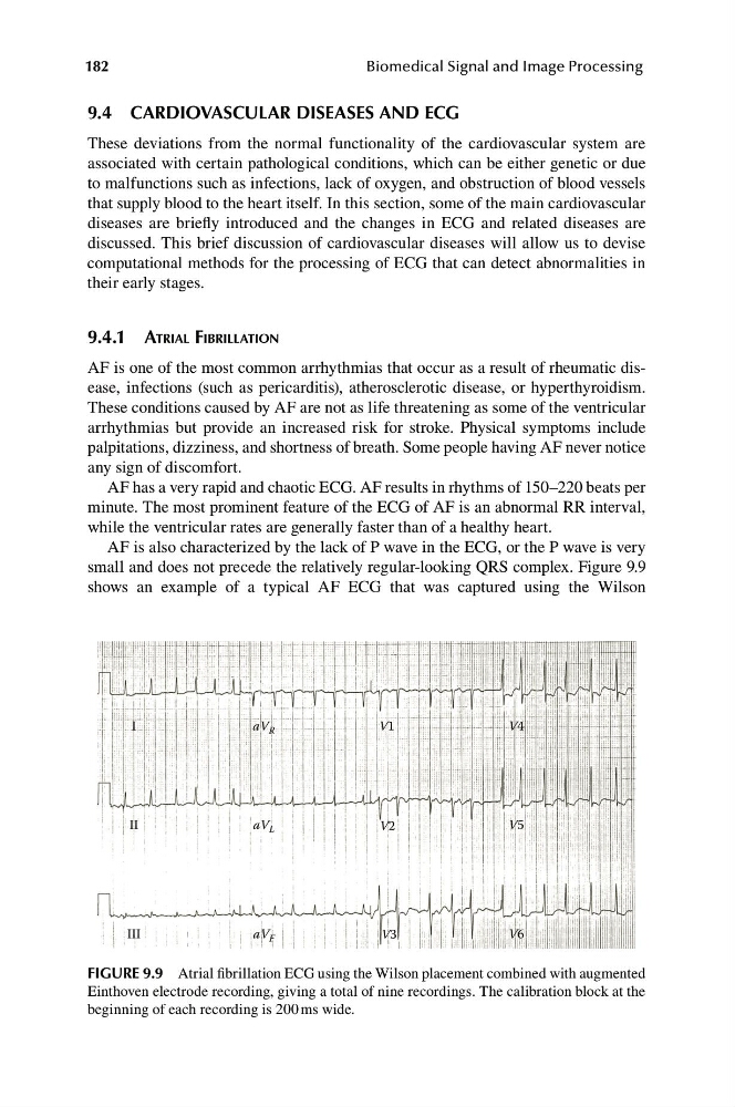
AF has a very rapid and chaotic ECG. AF results in rhythms of 150–220 beats per
minute. The most prominent feature of the ECG of AF is an abnormal RR interval,
while the ventricular rates are generally faster than of a healthy heart.
AF is also characterized by the lack of P wave in the ECG, or the P wave is very
small and does not precede the relatively regular-looking QRS complex. Figure 9.9
shows an example of a typical AF ECG that was captured using the Wilson
I
aV R
V1
V4
II
aV L
V2
V5
III
aV F
V3
V6
FIGURE 9.9 Atrial fibrillation ECG using the Wilson placement combined with augmented
Einthoven electrode recording, giving a total of nine recordings. The calibration block at the
beginning of each recording is 200 ms wide.
Biomedical Signal and Image Processing
9.4 CARDIOVASCULAR DISEASES AND ECG
These deviations from the normal functionality of the cardiovascular system are
associated with certain pathological conditions, which can be either genetic or due
to malfunctions such as infections, lack of oxygen, and obstruction of blood vessels
that supply blood to the heart itself. In this section, some of the main cardiovascular
diseases are briefly introduced and the changes in ECG and related diseases are
discussed. This brief discussion of cardiovascular diseases will allow us to devise
computational methods for the processing of ECG that can detect abnormalities in
their early stages.
9.4.1 ATRIAL FIBRILLATION
AF is one of the most common arrhythmias that occur as a result of rheumatic disease, infections (such as pericarditis), atherosclerotic disease, or hyperthyroidism.
These conditions caused by AF are not as life threatening as some of the ventricular
arrhythmias but provide an increased risk for stroke. Physical symptoms include
palpitations, dizziness, and shortness of breath. Some people having AF never notice
any sign of discomfort.
AF has a very rapid and chaotic ECG. AF results in rhythms of 150–220 beats per
minute. The most prominent feature of the ECG of AF is an abnormal RR interval,
while the ventricular rates are generally faster than of a healthy heart.
AF is also characterized by the lack of P wave in the ECG, or the P wave is very
small and does not precede the relatively regular-looking QRS complex. Figure 9.9
shows an example of a typical AF ECG that was captured using the Wilson
I
aV R
V1
V4
II
aV L
V2
V5
III
aV F
V3
V6
FIGURE 9.9 Atrial fibrillation ECG using the Wilson placement combined with augmented
Einthoven electrode recording, giving a total of nine recordings. The calibration block at the
beginning of each recording is 200 ms wide.