

172
Biomedical Signal and Image Processing
Pulmonary
vein
Aorta
LA
RV
RA
LV
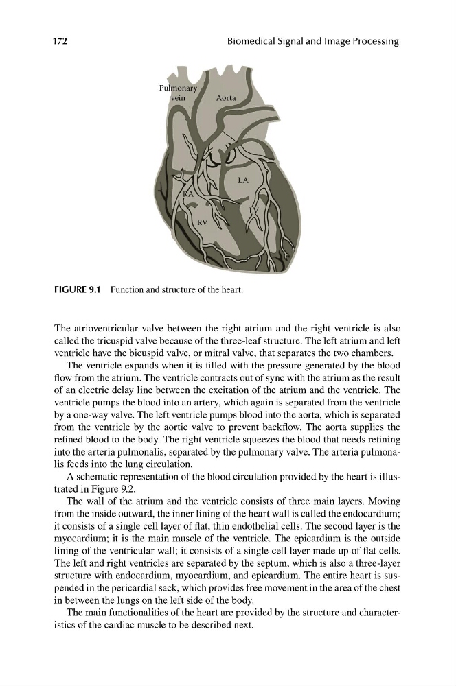
FIGURE 9.1 Function and structure of the heart.
The atrioventricular valve between the right atrium and the right ventricle is also
called the tricuspid valve because of the three-leaf structure. The left atrium and left
ventricle have the bicuspid valve, or mitral valve, that separates the two chambers.
The ventricle expands when it is filled with the pressure generated by the blood
flow from the atrium. The ventricle contracts out of sync with the atrium as the result
of an electric delay line between the excitation of the atrium and the ventricle. The
ventricle pumps the blood into an artery, which again is separated from the ventricle
by a one-way valve. The left ventricle pumps blood into the aorta, which is separated
from the ventricle by the aortic valve to prevent backflow. The aorta supplies the
refined blood to the body. The right ventricle squeezes the blood that needs refining
into the arteria pulmonalis, separated by the pulmonary valve. The arteria pulmonalis feeds into the lung circulation.
A schematic representation of the blood circulation provided by the heart is illustrated in Figure 9.2.
The wall of the atrium and the ventricle consists of three main layers. Moving
from the inside outward, the inner lining of the heart wall is called the endocardium;
it consists of a single cell layer of flat, thin endothelial cells. The second layer is the
myocardium; it is the main muscle of the ventricle. The epicardium is the outside
lining of the ventricular wall; it consists of a single cell layer made up of flat cells.
The left and right ventricles are separated by the septum, which is also a three-layer
structure with endocardium, myocardium, and epicardium. The entire heart is suspended in the pericardial sack, which provides free movement in the area of the chest
in between the lungs on the left side of the body.
The main functionalities of the heart are provided by the structure and characteristics of the cardiac muscle to be described next.
Biomedical Signal and Image Processing
Pulmonary
vein
Aorta
LA
RV
RA
LV
FIGURE 9.1 Function and structure of the heart.
The atrioventricular valve between the right atrium and the right ventricle is also
called the tricuspid valve because of the three-leaf structure. The left atrium and left
ventricle have the bicuspid valve, or mitral valve, that separates the two chambers.
The ventricle expands when it is filled with the pressure generated by the blood
flow from the atrium. The ventricle contracts out of sync with the atrium as the result
of an electric delay line between the excitation of the atrium and the ventricle. The
ventricle pumps the blood into an artery, which again is separated from the ventricle
by a one-way valve. The left ventricle pumps blood into the aorta, which is separated
from the ventricle by the aortic valve to prevent backflow. The aorta supplies the
refined blood to the body. The right ventricle squeezes the blood that needs refining
into the arteria pulmonalis, separated by the pulmonary valve. The arteria pulmonalis feeds into the lung circulation.
A schematic representation of the blood circulation provided by the heart is illustrated in Figure 9.2.
The wall of the atrium and the ventricle consists of three main layers. Moving
from the inside outward, the inner lining of the heart wall is called the endocardium;
it consists of a single cell layer of flat, thin endothelial cells. The second layer is the
myocardium; it is the main muscle of the ventricle. The epicardium is the outside
lining of the ventricular wall; it consists of a single cell layer made up of flat cells.
The left and right ventricles are separated by the septum, which is also a three-layer
structure with endocardium, myocardium, and epicardium. The entire heart is suspended in the pericardial sack, which provides free movement in the area of the chest
in between the lungs on the left side of the body.
The main functionalities of the heart are provided by the structure and characteristics of the cardiac muscle to be described next.